Skip to content
NBOME Surgery COMAT & Level 2-CE NBME Surgery Shelf Exam & Step 2 CK

Surgery COMAT Command Center Surgery Shelf Exam Command Center

General surgery, perioperative medicine, surgical acute abdomen, trauma ATLS protocols, and post-operative complications for NBOME Surgery COMAT and NBME Surgery Shelf exams. Synthesizes 300+ board-curated questions paired with 3 core surgical pathways.

72 Cards Extracted 3 High-Yield Protocols 4 Osteopathic Rules 4 High-Yield Pearls
Section 01 • Osteopathic Principles

"Don't Miss" COMAT Osteopathic Pearls

High-frequency somatic dysfunctions, viscerosomatics, autonomic reflexes, and treatment rules

Tested heavily on NBOME Shelf

Post-Operative Ileus & Autonomic Inhibition

Peritoneal irritation and bowel manipulation induce hyperactive sympathetic firing via the celiac (T5–T9), superior mesenteric (T10–T11), and inferior mesenteric (T12–L2) collateral ganglia, paralyzing bowel peristalsis. Gentle paraspinal rib raising and inhibitory sacral rocking (S2–S4 pelvic splanchnics) normalize sympathetic outflow, activate parasympathetic propulsive peristalsis, accelerate return of bowel sounds and flatus, and minimize opioid requirements.

Surgical Chapman Reflex Points for Acute Abdomen

Acute Appendicitis: Tip of right 12th rib anteriorly; between T11–T12 transverse processes on the right posteriorly. Acute Cholecystitis: Right 6th intercostal space between midclavicular and axillary lines; between T6–T7 spinous processes on the right. Pancreatitis: Right 7th intercostal space anteriorly; between T7–T8 on the right posteriorly. Colon/Bowel: Outer aspect of the iliotibial band from trochanter to knee.

Thoracotomy / Laparotomy Rib Mechanics & Post-Op Splinting

Subcostal or upper midline laparotomy incisions cause severe abdominal wall guarding and exhalation somatic dysfunctions of the inferior rib cage (ribs 6–10). This mechanical restriction dramatically reduces tidal volume, precipitating early post-operative atelectasis. Treating thoracic cage somatic dysfunctions and freeing Sibson's fascia (thoracic inlet) restores thoracic excursion, optimizes lymphatic return, and reduces pulmonary complication rates.

OMT Contraindications in Surgical Inpatients

Absolute Contraindications: Direct pressure or thrusting techniques over acute surgical wounds, abdominal incisions with suspected fascial dehiscence, acute unreduced fractures, deep venous thrombosis (vigorous lower extremity lymphatic pumps risk fatal pulmonary embolism), and acute peritonitis/free air perforation before exploratory laparotomy. Relative Contraindications: Mild local tissue edema, localized incisional discomfort away from the treatment site.

Section 01 • Clinical Foundations

"Don't Miss" Surgery Clinical Pearls & Shelf Traps

High-frequency diagnostic pitfalls, gold-standard criteria, and next-best-step clinical rules

Tested heavily on NBME Shelf & Step 2 CK

Post-Operative Fever Timeline: The 5 W's

Day 1–2: Wind (Atelectasis due to hypoventilation/splinting; treat with incentive spirometry and ambulation; not an infection). Day 3: Water (Urinary tract infection; inspect Foley catheter, remove if possible, check urinalysis/culture). Day 5: Wound (Surgical site infection; inspect incision for erythema/fluctuance; superficial open and pack, fascial dehiscence requires immediate OR). Day 7: Walking (Deep vein thrombosis / Pulmonary embolism; obtain venous duplex ultrasound or CT pulmonary angiography). Day 10+: Wonder drugs / Deep abscess (drug-induced fever [heparin, beta-lactams] or deep intra-abdominal/pelvic abscess; obtain CT scan with IV and oral contrast for percutaneous drainage).

Acute Abdomen Triage & Immediate Laparotomy Indications

Emergency surgical exploratory laparotomy is indicated without delay for: 1. Peritoneal signs on physical examination (involuntary guarding, rigid board-like abdomen, percussion tenderness); 2. Pneumoperitoneum (free air beneath the diaphragms on upright chest radiograph or CT); 3. Hemodynamically unstable trauma patient with free intra-abdominal fluid on FAST exam; 4. Complete closed-loop small bowel obstruction or bowel strangulation with systemic toxicity, peritonitis, or pneumatosis intestinalis.

Pre-Operative Cardiac Risk Stratification (Revised Cardiac Risk Index - RCRI)

Six independent clinical predictors of perioperative major cardiac complications: 1. High-risk surgery (intraperitoneal, intrathoracic, suprainguinal vascular); 2. Ischemic heart disease (history of MI, angina, positive stress test); 3. Congestive heart failure (history of HF, elevated BNP, S3 gallop); 4. Cerebrovascular disease (history of stroke or TIA); 5. Insulin-treated diabetes mellitus; 6. Pre-operative serum creatinine > 2.0 mg/dL. RCRI score >= 2 indicates high risk warranting pre-operative cardiology evaluation and non-invasive stress testing if functional capacity is < 4 METs.

Surgical Jaundice & Biliary Tree Differential

Charcot's Triad (RUQ pain, jaundice, fever) + Reynolds' Pentad (+ hypotension, altered mental status) indicates Acute Ascending Cholangitis from common bile duct obstruction (most commonly choledocholithiasis). Management: IV hydration, broad-spectrum antibiotics (pip/tazo), and emergent biliary decompression via ERCP (endoscopic retrograde cholangiopancreatography). Painless jaundice + palpable non-tender gallbladder (Courvoisier sign) indicates periampullary/pancreatic adenocarcinoma until proven otherwise.

Section 02 • Clinical Algorithms

Core Clinical Protocols & Diagnostic Trees

Step-by-step first-line management pathways, diagnostic thresholds, and pharmacological escalation

3 Diagnostic Algorithms

Acute Abdomen & Peritonitis Surgical Decision Tree

  1. 1. Primary Assessment: Assess hemodynamic stability (vitals, orthostatics, GCS). Establish two large-bore 16G peripheral IV lines and administer isotonic crystalloids (30 mL/kg for sepsis/hypovolemia).
  2. 2. Physical Examination: Palpate for peritoneal signs (involuntary guarding, rebound tenderness, rigid abdomen, Rovsing/psoas/obturator signs for appendicitis; Murphy sign for cholecystitis).
  3. 3. Initial Diagnostic Workup: Obtain STAT CBC, comprehensive metabolic panel, serum lactate, lipase, coagulation panel, type and screen, urinalysis, and urine pregnancy test in all females of reproductive age.
  4. 4. Imaging: Order STAT Upright Chest X-ray and Abdominal Series (KUB) to assess for subdiaphragmatic free air (pneumoperitoneum) and air-fluid levels (small bowel obstruction).
  5. 5. Surgical Emergency: If subdiaphragmatic free air, bowel evisceration, or peritonitis with hemodynamic instability is identified -> Place NPO, place nasogastric (NG) tube to suction, administer broad-spectrum IV antibiotics (Cefepime + Metronidazole or Piperacillin-Tazobactam), call the OR, and transport patient directly for Exploratory Laparotomy.
  6. 6. Diagnostic Equivocal Cases: In stable patients without overt peritonitis, obtain STAT CT abdomen and pelvis with IV contrast (CT angiography if mesenteric ischemia suspected; RUQ ultrasound if biliary disease suspected).

ATLS Primary Survey & Massive Transfusion in Blunt/Penetrating Trauma

  1. 1. Airway with In-Line Cervical Spine Protection: Assess airway patency, phonation, and debris; place cervical collar; perform rapid sequence intubation with in-line stabilization if GCS <= 8 or respiratory failure.
  2. 2. Breathing & Ventilation: Auscultate breath sounds bilaterally. Identify and immediately treat immediate thoracic life threats: Needle thoracostomy (2nd ICS MCL or 5th ICS AAL) followed by tube thoracostomy (chest tube) for tension pneumothorax; 3-sided occlusive dressing for open pneumothorax.
  3. 3. Circulation & Hemorrhage Control: Apply direct manual pressure to active external bleeding; apply commercial tourniquets to extremities; place pelvic binder over greater trochanters for suspected open-book pelvic fracture.
  4. 4. Focused Assessment with Sonography for Trauma (E-FAST): Evaluate 4 acoustic windows (pericardial/subxiphoid, right upper quadrant/Morison's pouch, left upper quadrant/splenorenal, and suprapubic/pelvis).
  5. 5. Hemorrhagic Shock Protocol: If unstable (SBP < 90, HR > 120) and FAST is positive in blunt abdominal trauma, transport patient immediately to the Operating Room for Exploratory Laparotomy.
  6. 6. Massive Transfusion Activation: Initiate balanced 1:1:1 resuscitation (1 unit PRBC : 1 unit FFP : 1 unit Platelets); administer Tranexamic Acid (TXA 1 g IV bolus over 10 min, then 1 g infusion over 8 hr) within 3 hours of injury; prevent hypothermia, acidosis, and hypocalcemia.

Post-Operative Fever & Wound Dehiscence Management

  1. 1. Assess Chronologic Timing: Post-op Day 1–2 (Wind: atelectasis); Day 3 (Water: UTI); Day 5 (Wound: SSI); Day 7 (Walking: DVT/PE); Day 10+ (Wonder drugs or deep intra-abdominal abscess).
  2. 2. Incision Inspection: Remove surgical dressings and inspect wound edges for erythema, induration, purulent drainage, crepitus, or fluctuance.
  3. 3. Superficial vs. Fascial Dehiscence: Palpate wound integrity. If only skin/subcutaneous tissue separates with intact rectus fascia -> Superficial dehiscence; open incision, evacuate hematoma/pus, pack with moist saline gauze, allow healing by secondary intention.
  4. 4. Fascial Dehiscence / Evisceration: Profuse salmon-pink serosanguinous fluid drainage indicates ruptured deep abdominal wall fascia. Evisceration (loops of bowel protruding through wound) is an acute surgical emergency.
  5. 5. Evisceration Immediate Action: Immediately cover exposed bowel with sterile saline-soaked gauze, instruct patient to remain in low Fowler's position with knees flexed, keep strictly NPO, place NG tube, administer broad-spectrum IV antibiotics, and transport urgently to the Operating Room for emergency fascial closure.
  6. 6. Necrotizing Fasciitis: Dishwater-gray drainage, dishwater fluid, severe pain out of proportion to skin findings, crepitus, and systemic toxicity require emergent surgical debridement and triple IV antibiotics (Vancomycin + Piperacillin-Tazobactam + Clindamycin).
Section 03 • Clinical Chapters & Active Recall

High-Yield Clerkship Review by System

Read structured textbook-style disease summaries or test yourself with active-recall flashcards across Surgery clinical systems.

Showing 7 of 7 continuous textbook chapters
Chapter 1 • Acute Abdomen & GI Emergencies
18 min TOC

Acute Abdomen & Gastrointestinal Emergencies

Peritonitis, Appendicitis, Biliary Emergencies, Bowel Obstruction & Perforated Viscus

Clinical Overview & Board Focus

The evaluation of acute abdominal pain is the cornerstone of general surgery board examinations. Rapid distinction between medical causes, conditions requiring urgent evaluation, and surgical emergencies mandating immediate laparotomy determines clinical survival. Key competencies include localized vs. diffuse peritonitis, staging and management of appendicitis, the clinical continuum of gallstone disease, and differentiating mechanical obstruction from paralytic ileus.

1.1 Clinical Triage & Indications for Emergency Exploratory Laparotomy

The term 'acute abdomen' encompasses a spectrum of surgical, medical, and gynecological conditions characterized by acute, severe abdominal pain that may require urgent operative intervention. Pathophysiologically, pain originates as visceral pain conducted via unmyelinated C-fibers within splanchnic nerves responding to stretch, distention, or ischemia. Visceral pain is dull, crampy, poorly localized, and referred to the embryonic midline: foregut structures (stomach, duodenum, biliary tree, pancreas) refer to the epigastrium (T5–T9); midgut structures (small intestine, appendix, ascending and proximal transverse colon) refer to the periumbilical region (T10–T11); and hindgut structures (distal transverse, descending colon, rectum) refer to the hypogastrium (T12–L2).

As the disease process progresses to irritate the parietal peritoneum, somatic pain develops. Somatic pain is transmitted by myelinated A-delta fibers within somatic spinal nerves, producing sharp, intense, exquisitely localized pain accompanied by involuntary muscular guarding, percussion tenderness, and board-like abdominal rigidity. The presence of true involuntary guarding (spasm of the abdominal wall musculature that does not relax when the patient is distracted or breathes out) and rebound tenderness constitutes generalized peritonitis.

Emergency exploratory laparotomy is indicated without delay when peritonitis is identified in the setting of hemodynamic instability, free air beneath the diaphragm (pneumoperitoneum), complete closed-loop bowel obstruction, or evisceration. In hemodynamically stable patients without overt peritonitis, contrast-enhanced CT of the abdomen and pelvis remains the gold standard imaging modality, offering sensitivity exceeding 95% for acute appendicitis, diverticulitis, bowel obstruction, and intra-abdominal abscesses.
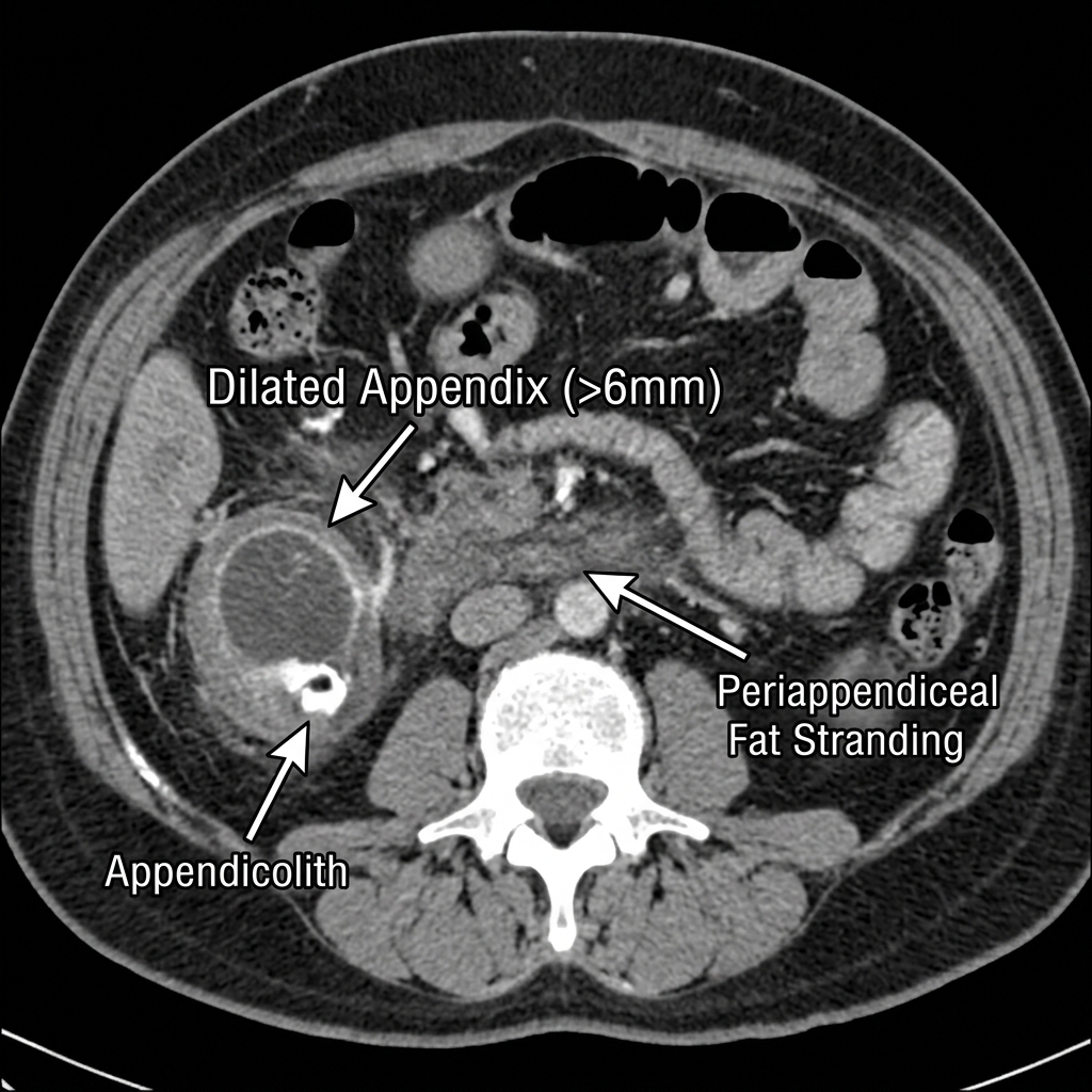
Acute Appendicitis on Contrast-Enhanced Abdominal CT
Axial contrast-enhanced CT demonstrating a dilated, fluid-filled appendix (> 6 mm outer diameter) with marked circumferential wall thickening, surrounding periappendiceal fat stranding, and a calcified appendicolith at the base. Appendectomy is the gold-standard definitive therapy.
Gastrointestinal Ganglia & Chapman Reflex Points
COMLEX & OPP Board Integration
Sympathetic innervation to the GI tract is partitioned across three prevertebral collateral ganglia: Celiac Ganglion (T5–T9) supplies foregut (stomach, liver, gallbladder, spleen, proximal duodenum); Superior Mesenteric Ganglion (T10–T11) supplies midgut (distal duodenum to proximal 2/3 transverse colon, appendix); and Inferior Mesenteric Ganglion (T12–L2) supplies hindgut (distal 1/3 transverse colon to rectum). Acute Appendicitis Chapman Point: Located on the tip of the right 12th rib anteriorly and between T11–T12 transverse processes on the right posteriorly. Acute inflammation produces profound right psoas hypertonicity and pelvic rotation.

1.2 The Biliary Tree Spectrum: From Cholelithiasis to Acute Ascending Cholangitis

Biliary tract diseases represent a continuous clinical spectrum dictated by the anatomical level and duration of gallstone impaction. Uncomplicated Cholelithiasis (Biliary Colic) occurs when a gallstone transiently impacts the cystic duct during gallbladder contraction stimulated by postprandial cholecystokinin (CCK). Patients present with severe, episodic, dull RUQ or epigastric pain radiating to the right infrascapular region, lasting 1 to 5 hours, resolving completely. Ultrasound reveals mobile gallstones without gallbladder wall thickening (< 3 mm) or pericholecystic fluid.

When persistent cystic duct obstruction causes sustained intraluminal distention, chemical inflammation, and secondary bacterial infection (E. coli, Klebsiella, Enterococcus), Acute Cholecystitis ensues. Pain becomes continuous, accompanied by low-grade fever, leukocytosis, and an arrest of inspiration upon deep RUQ palpation (Murphy sign). Ultrasound demonstrates gallstones, thickened gallbladder wall (> 3 mm), pericholecystic fluid, and a sonographic Murphy sign. If ultrasound is equivocal, HIDA scan (cholescintigraphy) is the most sensitive diagnostic test; failure to visualize the gallbladder after 4 hours confirms cystic duct obstruction. Management consists of NPO, IV fluid resuscitation, IV antibiotics, and laparoscopic cholecystectomy within 24 to 72 hours of admission.

Migration of a gallstone into the common bile duct produces Choledocholithiasis, characterized by biliary colic, jaundice (conjugated hyperbilirubinemia), and elevated alkaline phosphatase/GGT. If bacterial superinfection occurs proximal to the obstructed duct, life-threatening Acute Ascending Cholangitis develops. Patients exhibit Charcot's Triad (RUQ pain, jaundice, fever) or Reynolds' Pentad (adding hypotension and altered mental status, indicating septic shock). Management requires immediate aggressive IV resuscitation, broad-spectrum IV antibiotics (Piperacillin-Tazobactam), and emergent biliary decompression via Endoscopic Retrograde Cholangiopancreatography (ERCP).

Biliary Tree Pathology Comparison & Management Matrix
Clinical Matrix
Clinical EntityPathophysiologic MechanismHallmark FindingsDefinitive Management
Biliary ColicTransient cystic duct obstruction by gallstoneEpisodic RUQ pain < 6h; normal labs; thin wall on USElective outpatient laparoscopic cholecystectomy
Acute CholecystitisSustained cystic duct obstruction + inflammationConstant RUQ pain > 6h; Murphy sign; fever; wall > 3mmEarly laparoscopic cholecystectomy within 72 hr
CholedocholithiasisGallstone in common bile duct (CBD)Biliary pain + elevated direct bilirubin & alk phos; dilated CBDERCP stone extraction followed by cholecystectomy
Acute CholangitisInfected, obstructed CBD (bacterial stasis)Charcot triad (pain, jaundice, fever) / Reynolds pentadEmergent ERCP biliary decompression + IV antibiotics
Gallstone PancreatitisImpacted stone at ampulla of VaterEpigastric pain radiating to back + Lipase > 3x normalIV hydration; cholecystectomy prior to discharge
Board Trap — Painless Jaundice & Courvoisier Sign
High-Yield Board Trap & Alert
A palpable, non-tender, distended gallbladder in the setting of progressive painless jaundice (Courvoisier sign) is virtually never due to gallstone obstruction, as recurrent chronic cholecystitis produces a fibrotic, non-distensible gallbladder. Instead, it indicates malignant biliary obstruction, most commonly Adenocarcinoma of the Pancreatic Head or cholangiocarcinoma, until proven otherwise. Immediate evaluation requires a pancreatic-protocol CT scan.

1.3 Small Bowel Obstruction, Volvulus & Visceral Perforation

Small Bowel Obstruction (SBO) is among the most frequent indications for emergent general surgery admission. The leading etiology in patients with prior abdominal surgery is intra-abdominal adhesions (60–70%), followed by incarcerated groin or ventral hernias (15–20%) and malignancy. Patients present with crampy, colicky periumbilical abdominal pain, obstipation, nausea, and bilious vomiting. Early in the disease course, physical examination reveals hyperactive, high-pitched 'tinkling' bowel sounds; with progressive distention and vascular compromise, bowel sounds become absent. Abdominal radiographs demonstrate dilated loops of small bowel (> 3 cm) arranged in a 'stepladder' pattern with multiple air-fluid levels and a paucity of colonic gas.

Initial management of uncomplicated SBO is non-operative: strict NPO, nasogastric (NG) tube decompression to low intermittent suction, aggressive isotonic IV crystalloid resuscitation to replace third-spaced fluid, and monitoring of electrolyte panels. More than 70% of adhesive partial SBOs resolve with conservative therapy within 48 to 72 hours. However, emergency surgical exploration (laparotomy) is mandatory if signs of strangulation or closed-loop obstruction develop: fever, tachycardia, localized peritonitis, leukocytosis, metabolic lactic acidosis, or pneumatosis intestinalis on CT.

Visceral perforation manifests as sudden, catastrophic, severe abdominal pain resulting from chemical or bacterial peritonitis. Peptic ulcer perforation (most commonly anterior duodenal ulcer) releases gastric acid and bile into the peritoneal cavity, producing subdiaphragmatic free air (pneumoperitoneum) visible as a thin crescent beneath the right hemidiaphragm on an upright chest radiograph. Immediate management comprises NPO, NG tube suction, IV PPI, broad-spectrum IV antibiotics, and emergent laparotomy with omental patch closure (Graham patch).
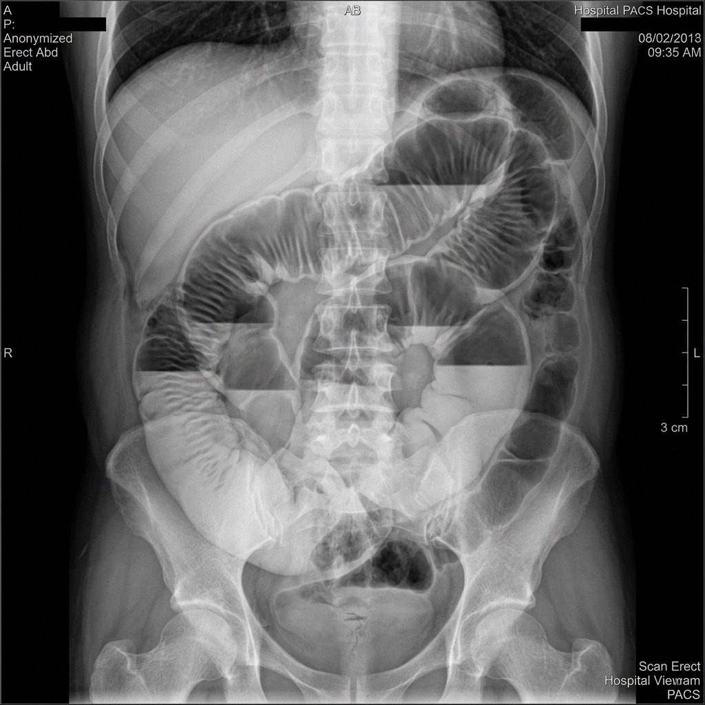
Small Bowel Obstruction on Abdominal Radiograph
Upright abdominal radiograph demonstrating hallmark multiple dilated small bowel loops with prominent plicae circulares (valvulae conniventes) crossing the entire width of the lumen and distinct air-fluid levels in an adhesive SBO.

Chapter 1 Quick-Check Self-Assessment

Test your clinical reasoning before moving to the next chapter
2 Vignettes
Board Vignette #1 Single Best Answer
A 45-year-old woman with a history of open appendectomy presents with obstipation, bilious vomiting, and crampy abdominal pain for 24 hours. Exam reveals distended abdomen with high-pitched tinkling sounds, no peritoneal signs. Vitals: BP 124/80, HR 88, T 37.0°C. What is the initial next step in management?
Board Vignette #2 Single Best Answer
A 67-year-old male smoker with hypertension presents with sudden, catastrophic epigastric pain that rapidly became diffuse. Abdomen is rigid and board-like with severe rebound tenderness. Upright CXR reveals crescentic air beneath the right diaphragm. What is the most appropriate next step?
Estimated study time: 18 min
Chapter 2 • Pre-Operative & Perioperative Care
16 min TOC

Pre-Operative Risk Stratification & Perioperative Care

Cardiopulmonary Risk Indices, Medication Adjustments, Prophylaxis & Fluid Balance

Clinical Overview & Board Focus

Meticulous pre-operative evaluation optimizes chronic comorbidities, identifies occult organ dysfunction, and substantially reduces surgical morbidity and mortality. Board questions emphasize the Revised Cardiac Risk Index (RCRI), functional capacity assessment (METs), perioperative antithrombotic management, glycemic control, prophylactic antimicrobial timing, and venous thromboembolism prevention protocols.

2.1 Cardiac & Pulmonary Risk Assessment

Cardiac complications (myocardial infarction, acute heart failure, malignant dysrhythmias) represent the foremost cause of perioperative mortality following non-cardiac surgery. The Revised Cardiac Risk Index (RCRI / Lee Index) assesses six independent clinical predictors of major adverse cardiac events: 1. High-risk surgical procedure (intraperitoneal, intrathoracic, or suprainguinal vascular); 2. History of ischemic heart disease (prior MI, positive exercise stress test, current angina, or Q waves); 3. History of congestive heart failure; 4. History of cerebrovascular disease (prior stroke or TIA); 5. Insulin-treated diabetes mellitus; and 6. Pre-operative serum creatinine > 2.0 mg/dL. Patients with an RCRI score >= 2 (moderate to high risk) require formal evaluation of functional capacity.

Functional capacity is quantified in Metabolic Equivalents (METs). The ability to climb two flights of stairs, walk up a hill, or run a short distance without stopping corresponds to >= 4 METs, indicating adequate physiological cardiopulmonary reserve. Patients with >= 4 METs can generally proceed directly to elective surgery without further cardiac testing, even in the presence of risk factors. Conversely, patients with poor functional capacity (< 4 METs) or unknown reserve who have elevated cardiac risk warrant non-invasive pharmacological stress testing (dobutamine stress echocardiography or myocardial perfusion imaging) prior to elective high-risk surgery.

Pulmonary risk factors rival cardiac complications in frequency. Significant risk factors include active tobacco smoking, chronic obstructive pulmonary disease (COPD), obstructive sleep apnea (OSA), age > 65, and planned surgical site close to the diaphragm (upper abdominal and thoracic incisions). Smoking cessation: Patients must stop smoking at least 4 to 8 weeks prior to elective surgery to demonstrate significant reductions in post-operative pulmonary complications and wound infections. Smoking cessation within 2 weeks prior to surgery may transiently increase bronchospasm and sputum volume, but is still encouraged to reduce carboxyhemoglobin levels within 24 to 48 hours.

Pre-Operative Rib Raising & Autonomic Homeostasis
COMLEX & OPP Board Integration
Pre-operative anxiety and physiological stress trigger systemic hypersympathetic arousal via thoracic chain ganglia (T1–T5 to heart/lungs; T5–L2 to abdominal viscera). Performing gentle rib raising along the costovertebral articulations and suboccipital release prior to surgery normalizes sympathetic outflow, enhances baseline vagal tone, and prevents intra-operative hypertensive spikes during anesthetic induction.

2.2 Perioperative Antithrombotic & Medication Management

Managing chronic antiplatelet and anticoagulant medications requires balancing the catastrophic risk of surgical hemorrhage against thromboembolism. In patients with coronary artery stents, elective non-cardiac surgery must be delayed whenever possible: at least 30 days following bare-metal stent (BMS) placement, and ideally 6 months (minimum 3 months) following drug-eluting stent (DES) placement to minimize stent thrombosis. When surgery cannot be delayed, dual antiplatelet therapy should be maintained if feasible, or P2Y12 inhibitors (Clopidogrel, Ticagrelor) held for 5 days (Prasugrel for 7 days) while continuing Aspirin.

Warfarin Management: Warfarin is held for 5 days prior to surgery to allow the International Normalized Ratio (INR) to normalize (< 1.5). In patients with high risk of thromboembolism (mechanical mitral valve, mechanical aortic valve with risk factors, AFib with CHA2DS2-VASc >= 7, or VTE within the past 3 months), bridging anticoagulation with therapeutic Low-Molecular-Weight Heparin (LMWH, enoxaparin 1 mg/kg SQ q12h) is initiated once INR drops below 2.0. The last dose of therapeutic LMWH is administered 24 hours prior to surgery. Direct Oral Anticoagulants (DOACs: Apixaban, Rivaroxaban) are held for 48 hours prior to high-bleeding-risk surgery without heparin bridging.

Chronic Corticosteroids: Patients taking > 5 mg daily of prednisone (or equivalent) for > 3 weeks within the past year have suppressed hypothalamic-pituitary-adrenal (HPA) axis reserve. Without perioperative supplemental steroids, the physiological stress of surgery precipitates acute secondary adrenal crisis (refractory hypotension, hypoglycemia). Patients undergoing moderate-to-high stress surgery require stress-dose hydrocortisone (100 mg IV prior to incision, followed by 50 mg IV q8h for 24 hours, then rapidly tapered).

Perioperative Medication Adjustment & Holding Guidelines
Clinical Matrix
Medication ClassPre-Operative Holding IntervalRationale / High-Yield Board Rule
WarfarinHold 5 days prior; check INR day of surgeryNormalize INR < 1.5; bridge with therapeutic LMWH if high VTE/valve risk
DOACs (Apixaban, Rivaroxaban)Hold 48 hours prior (72h if renal impairment)Rapid clearance; bridging is NOT required
Clopidogrel / TicagrelorHold 5 days prior (Prasugrel 7 days)Irreversible platelet inhibition; continue Aspirin if stent present
ACE Inhibitors / ARBsHold morning of surgery (24 hours)Prevents refractory vasoplegic hypotension during general anesthetic induction
Oral Hypoglycemics (Metformin)Hold 24–48 hours prior to surgeryMetformin held due to risk of lactic acidosis; SGLT2i held 3–4 days (euglycemic DKA)
Basal InsulinReduce to 50–75% of normal dose on morningPrevents ketogenesis while NPO; monitor blood glucose q2–4h

2.3 Surgical Antimicrobial Prophylaxis & Fluid Replacement

Antimicrobial prophylaxis aims to establish bactericidal concentrations in serum and tissues at the exact moment of skin incision. The gold-standard agent for clean-contaminated general surgical procedures (cholecystectomy, gastrectomy, bowel resection) is Cefazolin (2 g IV for patients < 120 kg, 3 g for >= 120 kg). Prophylactic antibiotics must be administered within 60 minutes prior to surgical incision (120 minutes for vancomycin or fluoroquinolones). Intra-operative redosing is mandatory if surgery extends beyond 2 half-lives of the antibiotic (every 4 hours for cefazolin) or if blood loss exceeds 1,500 mL. Prophylaxis must be discontinued within 24 hours post-operatively.

Intra-operative fluid management adheres to the 4-2-1 rule for baseline maintenance: 4 mL/kg/hr for the first 10 kg, 2 mL/kg/hr for the second 10 kg, and 1 mL/kg/hr for each remaining kg. In addition, third-space fluid losses from surgical tissue exposure and evaporation must be replaced: 1–2 mL/kg/hr for minimal trauma (hernia repair); 3–4 mL/kg/hr for moderate trauma (cholecystectomy); and 6–8 mL/kg/hr for severe trauma (open laparotomy, pancreatectomy). Balanced crystalloids (Lactated Ringer's or Plasma-Lyte) are preferred over 0.9% normal saline to prevent hyperchloremic metabolic acidosis.

Chapter 2 Quick-Check Self-Assessment

Test your clinical reasoning before moving to the next chapter
2 Vignettes
Board Vignette #1 Single Best Answer
A 68-year-old woman with a history of mechanical mitral valve replacement is scheduled for elective sigmoid resection for diverticulitis. Her current INR is 2.8 on warfarin. What is the most appropriate pre-operative anticoagulation management?
Board Vignette #2 Single Best Answer
What is the optimal timing for prophylactic intravenous cefazolin administration prior to skin incision in elective colorectal surgery?
Estimated study time: 16 min
Chapter 3 • Post-Operative Complications
17 min TOC

Post-Operative Complications & The 5 W's

Post-Op Fever Timeline, Surgical Site Infections, Wound Dehiscence & Oliguria

Clinical Overview & Board Focus

Mastery of post-operative complications requires an organized chronological framework. The classic '5 W's' (Wind, Water, Wound, Walking, Wonder drugs) guides evaluation of post-operative fever. Recognizing deep fascial dehiscence, differentiating superficial separation from surgical emergencies, and rapid diagnosis of life-threatening necrotizing fasciitis or malignant hyperthermia are high-frequency board concepts.

3.1 The Chronological Differential of Post-Operative Fever (The 5 W's)

Post-operative fever (temperature >= 38.3°C / 101.0°F) is a frequent clinical challenge. The chronological timing relative to the index operation provides the single most reliable diagnostic clue. Day 1 to 2 (Wind): Early fevers are almost uniformly due to Atelectasis, caused by hypoventilation, airway collapse, and diaphragmatic splinting from surgical pain and general anesthetics. Physical examination demonstrates shallow breathing and decreased breath sounds at the lung bases. Treatment relies on aggressive pulmonary toilet, early ambulation, and incentive spirometry; antibiotics are strictly unnecessary. If fever persists past day 3 with productive cough and infiltrates on chest X-ray, post-operative hospital-acquired pneumonia is diagnosed.

Day 3 (Water): Urinary Tract Infection (UTI) is the classic day 3 complication, typically associated with indwelling urinary catheters (CAUTI). Risk factors include prolonged catheterization, female sex, and diabetes. Diagnostic evaluation requires urinalysis (pyuria, positive leukocyte esterase, nitrites) and urine culture. Management consists of prompt removal of the Foley catheter and targeted antibiotic therapy.

Day 5 (Wound): Surgical Site Infection (SSI) typically manifests on post-operative day 5 to 7. Superficial SSIs involve skin and subcutaneous tissue, presenting with erythema, warmth, localized edema, and tenderness. First-line management is bedside wound opening, suture removal, evacuation of pus or hematoma, and wet-to-dry saline packing; antibiotics are indicated only if systemic toxicity or spreading cellulitis (> 5 cm) is present.

Day 7 (Walking): Deep Vein Thrombosis (DVT) and Pulmonary Embolism (PE) peak around post-operative day 7 due to stasis, endothelial injury, and perioperative hypercoagulability (Virchow triad). Unilateral calf swelling warrants duplex ultrasound; sudden dyspnea, pleuritic chest pain, or tachycardia warrants STAT CT Pulmonary Angiography (CTPA) and therapeutic anticoagulation.

Day 10+ (Wonder drugs & Deep Abscess): Late fevers result from Drug-induced fever (heparin, beta-lactam antibiotics, anticonvulsants) or occult Deep intra-abdominal/pelvic abscesses. Deep abscesses present with swinging fevers, leukocytosis, and deep localized tenderness. Abdominal CT with IV and oral contrast localizes the collection; percutaneous catheter drainage is the treatment of choice.

The Post-Operative Fever Timeline Matrix (The 5 W's)
Clinical Matrix
TimingClassic Etiology (The 5 W's)Pathophysiology & PresentationFirst-Line Clinical Management
Day 1–2Wind (Atelectasis)Alveolar collapse from shallow breathing, splinting, opioidsIncentive spirometry, early ambulation, analgesia; NO antibiotics
Day 3Water (UTI)Catheter-associated urinary tract infection (CAUTI)Remove Foley catheter, check urinalysis/culture, start antibiotics
Day 5Wound (Surgical Site Infection)Staphylococcus aureus, polymicrobial; erythema, fluctuanceOpen surgical wound, evacuate purulence, pack; antibiotics if cellulitis
Day 7Walking (DVT / PE)Thromboembolism from venous stasis, immobilizationDuplex ultrasound / CTPA; therapeutic anticoagulation (LMWH/heparin)
Day 10+Wonder drugs / Deep AbscessDrug fever (beta-lactams, heparin) or intra-abdominal abscessStop offending drugs; CT abdomen/pelvis + percutaneous drainage

3.2 Wound Dehiscence, Evisceration & Necrotizing Soft Tissue Infections

Wound disruption ranges from minor skin separation to life-threatening evisceration. Superficial Dehiscence involves separation of the skin and subcutaneous fat while the underlying musculofascial sheath remains intact. Management is conservative: open the skin, cleanse the wound, and pack with moist saline gauze to promote granulation tissue healing by secondary intention.

Deep Fascial Dehiscence involves rupture of the deep abdominal fascial closure line. The classic pathognomonic hallmark is sudden, copious discharge of clear, 'salmon-pink' serosanguinous peritoneal fluid soaking the surgical dressing, typically occurring on post-operative day 5 to 8. If untreated, intra-abdominal viscera herniate through the defect. Evisceration occurs when loops of bowel or omentum protrude through the disrupted abdominal incision. Evisceration is a surgical emergency: immediately cover the exposed bowel with sterile, saline-moistened gauze, place the patient in low Fowler's position with knees flexed to minimize abdominal wall tension, keep strictly NPO, place an NG tube to suction, administer IV broad-spectrum antibiotics, and transport immediately to the operating room for emergency fascial closure.

Necrotizing Soft Tissue Infection (Necrotizing Fasciitis): A rapidly progressive, fulminant infection of the deep fascia and subcutaneous tissues. Classic hallmarks include pain out of proportion to physical exam findings, dishwater-gray fluid discharge, soft tissue crepitus (subcutaneous gas), and rapid systemic toxicity (fever, tachycardia, hypotension). Treatment demands emergent, radical surgical debridement of all necrotic tissue back to bleeding viable fascia, combined with triple IV antimicrobial coverage: Vancomycin + Piperacillin-Tazobactam + Clindamycin (to shut down streptococcal protein and exotoxin synthesis).

Board Trap — Fascial Dehiscence vs. Superficial Separation
High-Yield Board Trap & Alert
Serosanguinous drainage that is pink, thin, and continuous ('salmon-pink fluid') indicates disruption of the deep abdominal fascia, NOT simple seroma or superficial wound separation. Never dismiss copious pink drainage as a benign seroma—probe the wound under sterile conditions to evaluate fascial integrity, and prepare for emergency operative closure if fascial defect is detected.

3.3 Post-Operative Oliguria & Malignant Hyperthermia

Post-operative oliguria is defined as urine output < 0.5 mL/kg/hr for 2 consecutive hours. The initial step is always to inspect the Foley catheter for mechanical kinks or blood clot obstruction and irrigate the catheter. If catheter patency is confirmed, distinguish prerenal azotemia (hypovolemia, third-spacing, bleeding) from intrinsic renal failure (acute tubular necrosis from prolonged hypotension or nephrotoxins). In patients without signs of fluid overload or heart failure, administer an initial fluid bolus (500 to 1,000 mL of isotonic crystalloid). If urine output responds promptly, prerenal hypovolemia is confirmed. If oliguria persists, check BUN, serum creatinine, urine electrolytes, and fractional excretion of sodium (FeNa < 1% indicates prerenal; FeNa > 2% indicates ATN).

Malignant Hyperthermia (MH): An inherited autosomal dominant pharmacogenetic disorder of the skeletal muscle ryanodine receptor (RYR1 gene). Exposure to triggering agents—all volatile halogenated inhalational anesthetics (halothane, isoflurane, sevoflurane, desflurane) and the depolarizing muscle relaxant succinylcholine—causes uncontrolled release of calcium from the sarcoplasmic reticulum into the myoplasm. Unchecked cellular metabolism produces masseter muscle spasm, severe hypercarbia (abrupt rise in end-tidal CO2 refractory to hyperventilation), tachycardia, generalized muscle rigidity, rhabdomyolysis, and hyperthermia exceeding 41°C. Treatment: 1. Immediately discontinue triggering agents; 2. Hyperventilate with 100% O2; 3. Administer IV Dantrolene Sodium (2.5 mg/kg IV bolus, repeated up to 10 mg/kg until symptoms abate); 4. Cool patient actively (cold IV saline, ice packs); and 5. Treat hyperkalemia aggressively with IV calcium gluconate, insulin, and glucose.

Post-Operative Ileus & Paraspinal Inhibition
COMLEX & OPP Board Integration
General anesthesia, retroperitoneal dissection, and surgical handling of bowel loops provoke intense reflex sympathetic outflow from the celiac, superior mesenteric, and inferior mesenteric ganglia (T5–L2), resulting in prolonged paralytic ileus. Performing paraspinal inhibitory pressure along T5–T9 (foregut) and T10–L2 (midgut/hindgut), along with sacral rocking (S2–S4 parasympathetics), dampens hypersympathetic tone, jump-starts colonic motility, and significantly shortens time to first flatus and bowel movement.

Chapter 3 Quick-Check Self-Assessment

Test your clinical reasoning before moving to the next chapter
2 Vignettes
Board Vignette #1 Single Best Answer
On post-operative day 6 following an exploratory laparotomy, a patient's wound dressing is saturated with copious thin, salmon-pink serosanguinous fluid. On palpation, loops of bowel are palpable beneath the separated skin. What is the immediate next step in management?
Board Vignette #2 Single Best Answer
Within 10 minutes of induction with sevoflurane and succinylcholine, a 24-year-old male develops masseter rigidity, sinus tachycardia (140 bpm), end-tidal CO2 rising to 80 mmHg, and temperature rising to 39.8°C. What is the mechanism of the definitive treatment agent?
Estimated study time: 17 min
Chapter 4 • Trauma Resuscitation & ATLS
18 min TOC

Trauma Resuscitation, Burns & ATLS Protocols

Primary Survey ABCDEs, FAST Ultrasound, Hemorrhagic Shock & Burn Resuscitation

Clinical Overview & Board Focus

Advanced Trauma Life Support (ATLS) establishes a standardized, stepwise methodology for trauma resuscitation. Clinical priorities dictate addressing immediate life threats in strict order: Airway, Breathing, Circulation, Disability, and Exposure (ABCDE). Differentiating shock etiologies with point-of-care ultrasound (E-FAST), massive transfusion protocols, and calculating burn fluid requirements via the Parkland formula are essential board competencies.

4.1 ATLS Primary Survey & Life-Threatening Thoracic Trauma

The ATLS Primary Survey prioritizes the identification and immediate reversal of conditions that kill trauma patients within minutes. A — Airway with in-line cervical spine stabilization: Assess vocalization and airway patency; clear blood and debris with suction. If Glasgow Coma Scale (GCS) is <= 8, severe facial trauma is present, or airway patency is compromised, perform emergency endotracheal intubation with continuous manual in-line cervical stabilization (avoid hyperextension of the neck). If orotracheal intubation fails due to severe maxillofacial trauma or massive hemorrhage, perform an emergency Surgical Cricothyroidotomy.

B — Breathing & Ventilation: Inspect chest excursion, auscultate bilaterally, and palpate for crepitus. Identify and immediately relieve immediate thoracic life threats: 1. Tension Pneumothorax: Tracheal deviation away from affected side, unilateral absent breath sounds, hyperresonance, and obstructive shock. Immediately perform needle thoracostomy (2nd intercostal space midclavicular line or 5th intercostal space anterior axillary line), followed promptly by placement of a formal chest tube (tube thoracostomy, 28–32 Fr). 2. Open Pneumothorax ('sucking chest wound'): Chest wall defect > 2/3 diameter of trachea. Immediately place a three-sided occlusive dressing (acting as a flutter valve) followed by tube thoracostomy at a separate site. 3. Massive Hemothorax: Rapid accumulation of > 1,500 mL of blood or > 200 mL/hr for 2 to 4 consecutive hours from a chest tube mandates emergent Exploratory Thoracotomy for surgical hemorrhage control.

3. Cardiac Tamponade: Penetrating trauma to the cardiac 'box' (midclavicular lines to epigastrium) resulting in hemopericardium. Classic Beck's Triad includes hypotension, jugular venous distention, and muffled heart sounds, accompanied by pulsus paradoxus (> 10 mmHg drop in SBP with inspiration). Bedside ultrasound (FAST) reveals pericardial effusion with diastolic right ventricular collapse. Emergent surgical relief (subxiphoid window, median sternotomy, or resuscitative thoracotomy) is required.

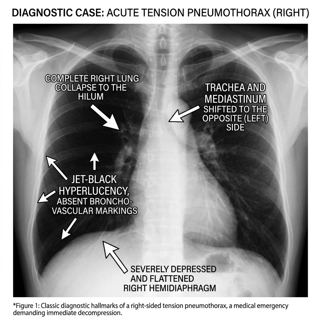
Tension Pneumothorax on Chest Radiograph
Supine chest radiograph illustrating total collapse of the right lung, depression of the right hemidiaphragm, and dramatic shift of the mediastinum and trachea toward the left. Treatment is immediate needle decompression without waiting for X-ray.
HVLA Contraindications in Trauma & Spinal Clearance
COMLEX & OPP Board Integration
High-Velocity Low-Amplitude (HVLA) thrust techniques are strictly contraindicated in any patient with acute trauma until complete anatomical and radiographic clearance of fractures, dislocations, ligamentous disruption, and spinal cord injury is documented. In cervical trauma, premature mobilization or manipulation can sever the spinal cord or lacerate the vertebral arteries.

4.2 Abdominal Trauma, E-FAST Ultrasound & Pelvic Fractures

C — Circulation with Hemorrhage Control: Hemorrhagic shock is the primary cause of preventable trauma death. Stop external bleeding with direct manual pressure or tourniquets. Assess hemodynamics (pulse, BP, capillary refill). Establish two large-bore (16-gauge or 14-gauge) peripheral IV lines or intraosseous (IO) access. In the presence of hemorrhagic shock, activate the Massive Transfusion Protocol (MTP), administering blood components in a balanced 1:1:1 ratio (1 unit Packed Red Blood Cells : 1 unit Fresh Frozen Plasma : 1 unit Platelets) to prevent dilutional coagulopathy. Administer Tranexamic Acid (TXA) (1 g IV over 10 min, then 1 g over 8 hr) within 3 hours of injury.

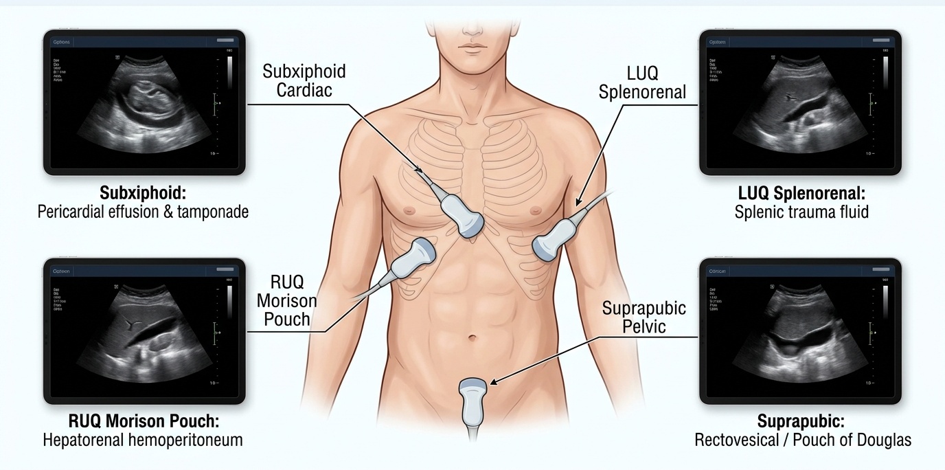
The Focused Assessment with Sonography for Trauma (FAST) evaluates four key acoustic windows for pathological fluid (blood): 1. Right Upper Quadrant (Morison's pouch): Space between liver and right kidney; the most sensitive dependent space in the supine abdomen; 2. Left Upper Quadrant (splenorenal recess): Space around spleen and left kidney; 3. Pelvis (Suprapubic view): Retrovesical pouch in males, rectouterine pouch (pouch of Douglas) in females; and 4. Pericardial (Subxiphoid view): Evaluates for pericardial effusion and tamponade. Critical Decision Rule: A hemodynamically unstable blunt trauma patient with a positive FAST must be transported directly to the operating room for Exploratory Laparotomy, NOT to the CT scanner.

Pelvic Ring Fractures: 'Open-book' pelvic fractures (disruption of pubic symphysis > 2.5 cm from anteroposterior compression) dramatically expand pelvic retroperitoneal volume, causing massive venous plexus bleeding. Immediate management is application of a commercial pelvic binder or bedsheet tightly wrapped around the level of the greater trochanters. If hypotension persists following pelvic binding and MTP, proceed to emergency pelvic angiography with embolization or preperitoneal pelvic packing.

E-FAST Ultrasound Acoustic Windows in Trauma
Point-of-care Focused Assessment with Sonography for Trauma (FAST) showing free fluid (anechoic black stripe) in Morison's pouch between the liver and right kidney in a patient with traumatic hemoperitoneum.

4.3 Severe Burns Management & The Parkland Formula

Thermal burns induce massive systemic capillary leak, evaporative fluid loss, and burn shock. Burn size is quantified using the Wallace Rule of Nines in adults: Head = 9%, Each Arm = 9%, Anterior Trunk = 18%, Posterior Trunk = 18%, Each Leg = 18%, and Perineum = 1%. Superficial (1st-degree) burns (erythema, pain, no blisters) are strictly excluded from TBSA calculations.

Fluid resuscitation for patients with >= 20% TBSA partial- and full-thickness burns is calculated using the Parkland Formula: 4 mL * Body Weight (kg) * % TBSA (2nd and 3rd degree) of Lactated Ringer's solution. 50% of the calculated total is administered in the first 8 hours calculated from the exact time of injury (not time of arrival), and the remaining 50% is infused over the subsequent 16 hours. Fluids must be continuously titrated to maintain target urine output: 0.5 to 1.0 mL/kg/hr in adults (1.0 to 2.0 mL/kg/hr in children).

Circumferential Full-Thickness Burns: Inelastic, leathery burn eschar creates a tourniquet effect. Circumferential chest burns restrict chest wall expansion, leading to hypoventilation and high peak airway pressures. Circumferential extremity burns compromise distal arterial flow, producing pulselessness and compartment syndrome. Treatment is immediate bedside Escharotomy (mid-lateral and mid-medial incisions through the entire thickness of the insensate eschar to relieve constriction).

Chapter 4 Quick-Check Self-Assessment

Test your clinical reasoning before moving to the next chapter

2 Vignettes
Board Vignette #1 Single Best Answer
A 32-year-old unrestrained driver involved in a high-speed MVC arrives with BP 78/42 mmHg, HR 135 bpm. E-FAST exam reveals free fluid in Morison's pouch and splenorenal recess. What is the most appropriate next step?
Board Vignette #2 Single Best Answer
An 80 kg male sustains 30% TBSA second- and third-degree flame burns in an explosion at 12:00 PM. He arrives at the trauma center at 2:00 PM. Using the Parkland formula, how much fluid should be administered between 2:00 PM and 8:00 PM (the remaining 6 hours of the first 8-hour window)?
Estimated study time: 18 min
Chapter 5 • Hernias & Abdominal Wall
15 min TOC

Abdominal Wall & Inguinal Hernias

Groin Anatomy, Direct vs. Indirect Inguinal, Femoral, Umbilical & Ventral Hernias

Clinical Overview & Board Focus

Groin and abdominal wall hernias represent the most common surgical pathology encountered in clinical practice. Board examinations heavily test precise anatomical boundaries (Hesselbach's triangle, femoral canal), differentiating direct from indirect inguinal hernias, identifying high-risk hernia variants (femoral, Richter, Littre), and recognizing acute strangulation requiring emergency intervention.

5.1 Inguinal & Femoral Hernia Comparative Anatomy

Groin hernias are anatomically classified according to their relationship to the inguinal ligament and the inferior epigastric vessels. Indirect Inguinal Hernias are the most common hernia in both men and women. They arise from a congenital failure of the processus vaginalis to obliterate, allowing peritoneal contents to pass through the deep (internal) inguinal ring, located lateral to the inferior epigastric vessels. The hernia sac travels down the inguinal canal alongside the structures of the spermatic cord (covered by the internal spermatic, cremasteric, and external spermatic fascias) and frequently extends into the scrotum or labium majus.

Direct Inguinal Hernias result from acquired progressive attenuation and weakness of the transversalis fascia (the floor of the inguinal canal) in older adult men due to chronic straining, heavy lifting, or COPD. They protrude directly anteriorly through Hesselbach's Triangle, located medial to the inferior epigastric vessels. The boundaries of Hesselbach's triangle are: 1. Inferior: Inguinal ligament (Poupart's ligament); 2. Medial: Lateral border of the Rectus abdominis muscle; and 3. Lateral / Superior: Inferior epigastric vessels. Direct hernias are covered only by the external spermatic fascia, do not travel into the scrotum, and have a wide neck with a very low risk of strangulation.

Femoral Hernias pass beneath the inguinal ligament through the femoral ring into the femoral canal, located medial to the femoral vein. Boundaries of the femoral ring are: anteriorly the inguinal ligament, posteriorly Cooper's ligament (pectineal ligament), medially the lacunar ligament (Gimbernat's ligament), and laterally the femoral vein. Femoral hernias occur predominantly in elderly multiparous women. Because the femoral ring is bounded by rigid, unyielding ligamentous structures, femoral hernias carry an exceptionally high risk of incarceration and strangulation (30–40%) and mandate prompt surgical repair upon diagnosis, even when completely asymptomatic.

Groin Hernia Comparative Anatomy & Clinical Distinctions
Clinical Matrix
Hernia TypeAnatomical Defect & LocationRelationship to Inf. Epigastric VesselsStrangulation Risk & Management
Indirect InguinalPatent processus vaginalis; deep inguinal ringLateral to inferior epigastric vesselsModerate; elective surgical repair with mesh
Direct InguinalWeakness of transversalis fascia (Hesselbach's)Medial to inferior epigastric vesselsLow; wide neck; elective repair if symptomatic
Femoral HerniaFemoral canal medial to femoral veinInferior to inguinal ligamentVERY HIGH (30–40%); urgent surgical repair mandatory
Board Trap — Femoral Hernia Incarceration Urgency
High-Yield Board Trap & Alert
Unlike direct inguinal hernias, which can be managed with watchful waiting if asymptomatic, Femoral Hernias must NEVER be managed expectantly. Due to the unyielding fibrous borders of the femoral ring (lacunar and Cooper's ligaments), up to 40% of femoral hernias present emergently with acute incarceration, bowel strangulation, and gangrene. Prompt elective repair is universally indicated upon discovery.

5.2 Hernia Staging, Rare Variants & Incisional Hernias

Hernias are clinically categorized into three functional stages: 1. Reducible: The hernia sac contents can be pushed back into the peritoneal cavity manually or spontaneously when the patient is supine; 2. Incarcerated: The hernia contents are permanently trapped within the defect due to adhesions or narrow neck, but vascular perfusion is intact; and 3. Strangulated: Vascular compromise of the trapped bowel wall, leading to arterial insufficiency, ischemic gangrene, and visceral perforation. Physical exam hallmarks of strangulation: Erythema of the overlying skin, severe localized tenderness, fever, leukocytosis, and tachycardia. Critical Rule: Forceful manual reduction (taxis) of a strangulated hernia is strictly contraindicated, as it risks reducing necrotic bowel back into the peritoneal cavity ('reduction en masse'), causing fatal peritonitis.

High-yield eponymous hernia variants tested on board exams include: Richter Hernia: Incarceration of only a portion of the antimesenteric wall of the intestine; causes gangrene and perforation without mechanical bowel obstruction. Littre Hernia: Hernia containing a Meckel's diverticulum. Amyand Hernia: Inguinal hernia sac containing the vermiform appendix. Spigelian Hernia: Protrusion through the Spigelian fascia along the semilunar line at the lateral edge of the rectus sheath, typically at or below the arcuate line.

Incisional & Ventral Hernias: Incisional hernias develop at prior laparotomy incision sites. The single most powerful independent risk factor is a prior post-operative surgical site infection (SSI), which impairs normal fascial collagen deposition. Additional risk factors include obesity, malnutrition, smoking, systemic corticosteroids, and technical surgical error (inadequate bite size or tension). Repair of incisional defects >= 2–3 cm requires tension-free placement of synthetic prosthetic mesh.

Chapter 5 Quick-Check Self-Assessment

Test your clinical reasoning before moving to the next chapter

2 Vignettes
Board Vignette #1 Single Best Answer
An 81-year-old woman presents with severe nausea, vomiting, and a painful, non-reducible mass in the right groin below the inguinal ligament, medial to the femoral artery pulsation. Overlying skin is warm and erythematous. What is the diagnosis and appropriate management?
Board Vignette #2 Single Best Answer
During laparoscopic groin hernia repair, an indirect inguinal hernia is identified. What is its anatomical relationship to the inferior epigastric vessels?
Estimated study time: 15 min
Chapter 6 • Vascular Surgery & Emergencies
16 min TOC

Vascular Surgery & Acute Vascular Emergencies

Abdominal Aortic Aneurysms, Aortic Dissection, Acute Limb Ischemia & Carotid Disease

Clinical Overview & Board Focus

Acute vascular emergencies demand rapid hemodynamic stabilization, accurate diagnostic imaging, and time-sensitive revascularization. Key board topics include screening, surveillance, and rupture of Abdominal Aortic Aneurysms (AAA); differentiating Stanford Type A vs. Type B Acute Aortic Dissection; the '6 P's' of Acute Arterial Limb Ischemia; and indications for Carotid Endarterectomy (CEA).

6.1 Abdominal Aortic Aneurysms (AAA) & Acute Aortic Dissection

An Abdominal Aortic Aneurysm (AAA) is defined as a permanent focal dilation of the abdominal aorta >= 3.0 cm, or > 50% increase compared to normal vessel diameter. The overwhelming majority (> 90%) are infrarenal, located between the renal arteries and the aortic bifurcation. Major risk factors include cigarette smoking (most potent modifiable risk factor), male sex, age > 65, atherosclerosis, and family history. Screening: The USPSTF recommends a one-time screening abdominal ultrasound for men aged 65 to 75 who have EVER smoked (>= 100 cigarettes).

Elective Repair Indications: The annual rupture risk rises exponentially with diameter. Elective surgical repair (via Endovascular Aneurysm Repair [EVAR] or open surgical grafting) is indicated when: 1. Aneurysm diameter reaches >= 5.5 cm in men or >= 5.0 cm in women; 2. Rapid expansion occurs (> 0.5 cm in 6 months or > 1.0 cm in 1 year); or 3. The aneurysm becomes symptomatic (tender on palpation). Ruptured AAA: Presents with the classic triad of sudden severe abdominal/flank/back pain, pulsatile abdominal mass, and hypotension/syncope. In an unstable patient with known or suspected AAA, transport immediately to the operating room for emergency repair without delaying for CT imaging.

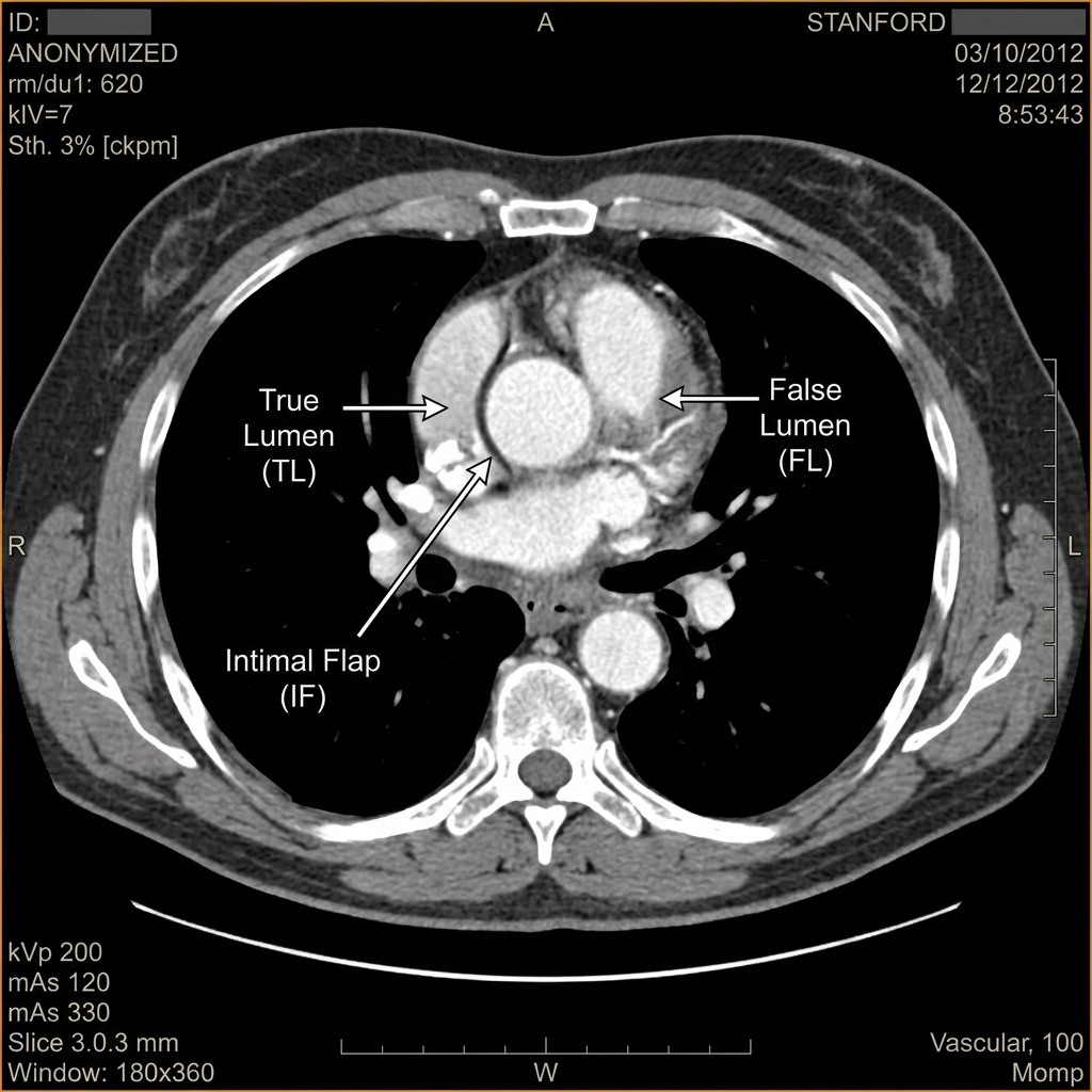
Acute Aortic Dissection: Initiated by a tear in the aortic intima, allowing systemic arterial blood under pulsatile pressure to cleave the media, creating a false lumen. Patients present with sudden-onset, catastrophic, 'ripping' or 'tearing' retrosternal or interscapular back pain. Physical examination reveals asymmetric peripheral pulses or blood pressure differential (> 20 mmHg) between arms, and a new diastolic decrescendo murmur of aortic regurgitation. Stanford Classification: Type A involves the ascending aorta (proximal to brachiocephalic artery) and is an immediate surgical emergency requiring open replacement of the ascending aorta. Type B involves only the descending aorta distal to the left subclavian artery; uncomplicated cases are managed medically with tight blood pressure and heart rate control (IV beta-blockers [Esmolol or Labetalol] targeting HR < 60 bpm and SBP 100–120 mmHg).

Acute Aortic Dissection on Contrast CT
Axial contrast-enhanced CT of the chest demonstrating a prominent intimal flap dividing the true and false lumens in a Stanford Type A aortic dissection involving the ascending thoracic aorta.
Board Trap — Stanford Type A vs. Type B Management
High-Yield Board Trap & Alert
Never manage Stanford Type A aortic dissection with medical therapy alone! Retrograde propagation of Type A dissection causes fatal cardiac tamponade (hemopericardium), acute coronary occlusion (causing inferior STEMI), or catastrophic acute aortic insufficiency. Emergent open cardiothoracic surgical repair is mandatory.

6.2 Acute Arterial Limb Ischemia & Compartment Syndrome

Acute Arterial Limb Ischemia is a sudden decrease in limb perfusion that threatens tissue viability. Etiologies divide into arterial embolism (80% from a cardiac source: atrial fibrillation, post-MI mural thrombus, or prosthetic valve) and in situ thrombosis over pre-existing atherosclerotic plaque. Embolic occlusion produces sudden, dramatic symptoms, most commonly lodging at the common femoral artery bifurcation. Presentation is remembered by the 6 P's: Pain (sudden, severe, distal), Pallor (waxy, cadaveric appearance), Poikilothermia (cold limb reflecting ambient temperature), Pulselessness (absent distal Doppler signals), Paresthesias (loss of light touch; early sign of nerve ischemia), and Paralysis (inability to move digits; late sign of irreversible neuromuscular necrosis).

Management is time-critical: irreversible muscle damage occurs within 4 to 6 hours of complete ischemia. Immediate intervention: administer an IV Unfractionated Heparin bolus (80 units/kg) followed by continuous infusion to halt clot propagation, and proceed immediately to emergency surgical revascularization (balloon catheter embolectomy via Fogarty catheter) or catheter-directed thrombolysis.

Ischemia-Reperfusion & Compartment Syndrome: Following restoration of arterial flow to a severely ischemic extremity, post-ischemic capillary endothelial leakage produces massive soft tissue edema within non-yielding fascial compartments. If intracompartmental pressure rises to within 30 mmHg of diastolic blood pressure, microvascular capillary perfusion stops. Hallmarks: Tense, 'woody' compartment firmness, excruciating pain on passive muscle stretch, and paresthesias. Emergency Treatment: Immediate bilateral double-incision four-compartment calf fasciotomy (releasing anterior, lateral, superficial posterior, and deep posterior compartments) to prevent permanent Volkmann ischemic contracture and amputation.

Chapter 6 Quick-Check Self-Assessment

Test your clinical reasoning before moving to the next chapter

2 Vignettes
Board Vignette #1 Single Best Answer
A 71-year-old male with atrial fibrillation presents with sudden, excruciating pain in his right leg. Exam shows a cold, pale right lower leg with absent dorsalis pedis and posterior tibial pulses. Light touch is decreased over the foot. What is the immediate first-line medical intervention?
Board Vignette #2 Single Best Answer
A 56-year-old man presents with tearing chest pain radiating to his back. SBP is 188/104 mmHg, HR is 112 bpm. CT scan confirms a Stanford Type B aortic dissection without evidence of malperfusion or rupture. What is the initial medical therapy of choice?
Estimated study time: 16 min
Chapter 7 • Surgical Oncology & Endocrine
17 min TOC

Surgical Oncology, Endocrine & Breast Surgery

Thyroid Nodules, Hyperparathyroidism, Adrenal Masses & Breast Cancer Management

Clinical Overview & Board Focus

Surgical oncology and endocrine surgery integrate surgical anatomy, histopathology, receptor biology, and targeted therapies. Board questions emphasize workup of solitary thyroid nodules, primary hyperparathyroidism localization and indications for parathyroidectomy, Multiple Endocrine Neoplasia (MEN) syndromes, pre-operative preparation of pheochromocytoma, and multimodality management of breast cancer.

7.1 Thyroid Nodule Workup & Primary Hyperparathyroidism

The initial step in evaluating a clinically detected solitary thyroid nodule is measuring serum TSH. If TSH is suppressed (low), obtain a Radionuclide Thyroid Iodine Uptake Scan. Hyperfunctioning ('hot') nodules are virtually never malignant and are managed medically, with radioactive iodine ablation, or with toxic adenoma excision. If serum TSH is normal or elevated, proceed to high-resolution Thyroid Ultrasonography. High-risk sonographic features include microcalcifications, hypoechogenicity, irregular or lobulated margins, absence of halo, and taller-than-wide shape. Fine-Needle Aspiration (FNA) Biopsy is indicated for nodules with suspicious sonographic features >= 1.0 cm, or >= 1.5 cm for intermediate features.

Thyroid Malignancies: 1. Papillary Thyroid Carcinoma (80%): Most common; excellent prognosis; microscopic Orphan Annie eye nuclei, nuclear grooves, and psammoma bodies; spreads via regional cervical lymphatics; treated with lobectomy or total thyroidectomy +/- radioactive iodine. 2. Follicular Thyroid Carcinoma (10–15%): Spreads hematogenously (to bone and lungs); FNA cannot distinguish benign adenoma from malignant carcinoma because diagnosis requires histological demonstration of capsular or vascular invasion. 3. Medullary Thyroid Carcinoma (5%): Arises from calcitonin-secreting parafollicular C-cells; amyloid stroma on histology; associated with MEN2A and MEN2B (RET proto-oncogene). 4. Anaplastic Thyroid Carcinoma (< 2%): Extremely aggressive; elderly patients presenting with rapidly enlarging, woody, fixed neck mass causing tracheal compression and dysphagia; dismal prognosis.

Primary Hyperparathyroidism (PHPT): Autonomous overproduction of parathyroid hormone (PTH) characterized by hypercalcemia, hypophosphatemia, and elevated intact PTH. Etiology: Solitary parathyroid adenoma (85%), four-gland hyperplasia (15%), or parathyroid carcinoma (< 1%). Pre-operative localization utilizes Technetium-99m Sestamibi scintigraphy or 4D-CT. Indications for surgical Parathyroidectomy in asymptomatic patients: 1. Age < 50; 2. Serum calcium > 1.0 mg/dL above normal; 3. Bone mineral density T-score < -2.5 (osteoporosis); or 4. eGFR < 60 mL/min or nephrolithiasis. Intra-operative PTH monitoring confirms cure (a > 50% drop in serum PTH 10 minutes following adenoma excision).

Cervical Fascial Mechanics & Post-Thyroidectomy Care
COMLEX & OPP Board Integration
The thyroid gland resides within the visceral compartment of the pretracheal fascia, anchored to the cricoid cartilage by the ligament of Berry. Upper thoracic and cervical somatic dysfunctions (OA, AA, C3–C5, and Rib 1) produce visceral fascial tension and lymphatic congestion in the neck. Following neck surgery, gentle indirect myofascial release and opening the thoracic inlet enhances lymphatic drainage and minimizes post-operative cervical hematoma tension.

7.2 Multiple Endocrine Neoplasia (MEN) & Pheochromocytoma

Multiple Endocrine Neoplasia (MEN) syndromes are autosomal dominant neuroendocrine disorders tested heavily on shelf exams. MEN 1 (Wermer Syndrome): Mutation in the *MEN1* gene encoding menin. Characterized by the 3 P's: Parathyroid hyperplasia (> 95%), Pituitary adenomas (prolactinoma), and Pancreatic neuroendocrine tumors (gastrinoma [Zollinger-Ellison], insulinoma, VIPoma).

MEN 2A: Mutation in the *RET proto-oncogene*. Characterized by: Medullary Thyroid Carcinoma (100%), Pheochromocytoma (50%), and Parathyroid hyperplasia (20–30%). MEN 2B: Also caused by *RET* mutations. Characterized by: Medullary Thyroid Carcinoma (very aggressive, occurring in infancy), Pheochromocytoma, Mucosal Neuromas (lips, tongue), and Marfanoid habitus (long limbs, joint laxity, but without lens dislocation or aortic aneurysm).

Pheochromocytoma: Catecholamine-secreting tumor of the adrenal medulla chromaffin cells. Presents with the classic triad of headache, diaphoresis, and tachycardia, along with episodic hypertension. Diagnosis: Elevated 24-hour urinary or plasma free fractionated metanephrines. Mandatory Pre-Operative Preparation: Initiate Alpha-Adrenergic Blockade FIRST (Phenoxybenzamine 10–14 days prior to surgery) to restore plasma volume and control blood pressure. Administer Beta-Blockers (Propranolol) ONLY AFTER adequate alpha-blockade has been established (2–3 days prior to surgery). Administering a beta-blocker first causes unopposed alpha-1 vasoconstriction, precipitating catastrophic hypertensive crisis, cerebral hemorrhage, and pulmonary edema.

Board Trap — Pheochromocytoma Alpha-Blockade Rule
High-Yield Board Trap & Alert
ALPHA BLOCKADE ALWAYS PRECEDES BETA BLOCKADE! This is one of the most frequently tested pharmacological rules in board exams. If an untreated pheochromocytoma patient receives a beta-blocker first, peripheral vasodilation is eliminated while alpha-1 vasoconstriction remains unchecked, causing massive arterial vasoconstriction and lethal hypertensive crisis.

7.3 Breast Cancer Surgical Evaluation & Management

Evaluation of a suspicious palpable breast mass or abnormal screening mammogram (BI-RADS 4 or 5) begins with diagnostic mammography and breast ultrasound, followed by core-needle biopsy for histological diagnosis and receptor testing (Estrogen Receptor [ER], Progesterone Receptor [PR], and Human Epidermal Growth Factor Receptor 2 [HER2/neu]).

Surgical Options for Early-Stage Disease: 1. Breast-Conserving Surgery (Lumpectomy / Partial Mastectomy): Complete surgical excision of the tumor with negative margins (no ink on tumor) followed by mandatory whole-breast adjuvant radiotherapy. Overall survival is identical to total mastectomy. Contraindications to lumpectomy: Multicentric disease (two or more separate tumors in different quadrants), widespread malignant-appearing microcalcifications, prior breast irradiation, active connective tissue disease (scleroderma), or persistent positive margins despite re-excision. 2. Total (Simple) Mastectomy: Entire breast tissue and nipple-areola complex removed, without axillary node dissection unless positive.

Axillary Staging: Sentinel Lymph Node Biopsy (SLNB) is standard of care for clinically node-negative (cN0) invasive breast cancer. Radioactive sulfur colloid and isosulfan blue dye are injected periareolarly; the first 1 to 3 draining lymph nodes are excised. If sentinel nodes are negative, formal axillary lymph node dissection (ALND) is avoided, preventing morbid lymphedema. Adjuvant Systemic Therapy: Postmenopausal women with ER/PR-positive tumors receive Aromatase Inhibitors (Anastrozole, Letrozole) for 5 to 10 years; premenopausal women receive Tamoxifen (selective estrogen receptor modulator [SERM]). Patients with HER2-positive tumors receive Trastuzumab (monoclonal antibody against HER2, with monitoring of cardiac ejection fraction).

Chapter 7 Quick-Check Self-Assessment

Test your clinical reasoning before moving to the next chapter

2 Vignettes
Board Vignette #1 Single Best Answer
A 42-year-old woman with a palpable 1.8 cm solitary thyroid nodule has a normal serum TSH. Ultrasound demonstrates a solid, hypoechoic nodule with internal microcalcifications and irregular margins. What is the most appropriate next step in management?
Board Vignette #2 Single Best Answer
A 36-year-old male with an adrenal mass and elevated urinary metanephrines is being prepared for laparoscopic adrenalectomy. What is the correct sequence of pre-operative pharmacological optimization?
Estimated study time: 17 min