Skip to content
NBOME Pediatrics COMAT & Level 2-CE NBME Pediatrics Shelf Exam & Step 2 CK

Pediatrics COMAT Command Center Pediatrics Shelf Exam Command Center

Comprehensive pediatric clerkship guide for NBOME Pediatrics COMAT and COMLEX Level 2-CE. Ingests 2,000+ pediatric cards covering developmental milestones, vaccination schedules, neonatal resuscitation, pediatric rashes, and pediatric OMM.

69 Cards Extracted 2 High-Yield Protocols 3 Osteopathic Rules 4 High-Yield Pearls
Section 01 • Osteopathic Principles

"Don't Miss" COMAT Osteopathic Pearls

High-frequency somatic dysfunctions, viscerosomatics, autonomic reflexes, and treatment rules

Tested heavily on NBOME Shelf

Neonatal Condylar Compression & Suckling Dysfunction

Birth trauma or prolonged labor can compress the occipital condyles against the atlas (C1), impinging the hypoglossal nerve (CN XII) within the hypoglossal canal. Manifests as poor suckling, tongue tremor, and feeding difficulties. Condylar decompression restores normal cranial base alignment and relieves nerve entrapment.

Recurrent Otitis Media & The Galbreath Technique

Otitis media is exacerbated by Eustachian tube dysfunction and lymphatic congestion. The Galbreath technique utilizes gentle inferomedial traction on the mandible to open the Eustachian tube and encourage lymphatic drainage through the pharyngeal plexus. Combined with thoracic pump and cervical lymphatic drainage.

Pediatric Torticollis & Plagiocephaly

Congenital muscular torticollis involves shortening of the sternocleidomastoid muscle (CN XI). Involves ipsilateral sidebending and contralateral rotation of the cranium. Evaluated for concurrent developmental dysplasia of the hip (DDH) and treated with gentle indirect myofascial release, cranial molding, and passive stretching.

Section 01 • Clinical Foundations

"Don't Miss" Pediatrics Clinical Pearls & Shelf Traps

High-frequency diagnostic pitfalls, gold-standard criteria, and next-best-step clinical rules

Tested heavily on NBME Shelf & Step 2 CK

Neonatal Hyperbilirubinemia: Physiologic vs. Pathologic

Pathologic Jaundice criteria (requires immediate diagnostic workup): Onset in the first 24 hours of life; direct (conjugated) bilirubin > 1.0 mg/dL (or > 20% of total); total bilirubin rising > 0.5 mg/dL/hr or > 5 mg/dL/day; or persisting > 2 weeks in term infants. Always plot total serum bilirubin on the hourly Bhutani nomogram to determine phototherapy or exchange transfusion thresholds (exchange indicated for TSB >= 25 mg/dL or signs of acute bilirubin encephalopathy/kernicterus). Breast milk jaundice peaks at week 2 due to beta-glucuronidase in milk; breastfeeding failure jaundice occurs in week 1 due to dehydration/inadequate intake.

Pediatric Stridor: Croup vs. Epiglottitis vs. Tracheitis

Croup (Laryngotracheobronchitis): Parainfluenza virus, age 6mo–3yr, 'barking' seal cough, inspiratory stridor, subglottic narrowing ('steeple sign' on AP neck X-ray); treatment is single-dose oral Dexamethasone (0.6 mg/kg) + nebulized Racemic Epinephrine if stridor at rest. Epiglottitis: Haemophilus influenzae type b, toxic appearance, rapid onset high fever, tripod position, drooling, absence of cough, 'thumbprint sign' on lateral neck X-ray; immediate emergency endotracheal intubation in operating room with ENT/anesthesia on standby.

Pediatric Limp & Hip Pain: Septic Arthritis vs. Transient Synovitis vs. SCFE

Kocher Criteria for Septic Arthritis: 1. Inability to bear weight, 2. Fever > 38.5°C (101.3°F), 3. ESR > 40 mm/hr, 4. WBC > 12,000/uL. If 4/4 criteria present = 99% probability -> emergent hip arthrocentesis (synovial WBC > 50,000/uL confirms). Transient Synovitis: Age 3–8, follows viral URI, afebrile/low fever, normal/mild labs, responds to NSAIDs. Slipped Capital Femoral Epiphysis (SCFE): Obese adolescent male (10–16 yr), dull hip/groin or referred knee pain, external rotation deformity on hip flexion; obtain bilateral frog-leg lateral X-rays ('ice cream falling off cone'); emergency non-weight-bearing and internal screw fixation.

Pediatric Dehydration: Clinical Staging & Rehydration Rules

Mild (3–5%): Normal vitals, moist mucosa, alert -> oral rehydration solution (ORS). Moderate (6–9%): Tachycardia, sunken fontanelle/eyes, dry mucosa, skin turgor delayed 2-3 sec, irritability -> ORS 50–100 mL/kg over 4 hours. Severe (>= 10%): Hypotension, lethargy/coma, capillary refill > 3 sec, cool mottled extremities, absent tears/urine -> emergent IV bolus of 20 mL/kg isotonic crystalloid (Normal Saline or Ringer's Lactate) over 10–20 minutes, repeated up to 3 times until peripheral perfusion restores.

Section 02 • Clinical Algorithms

Core Clinical Protocols & Diagnostic Trees

Step-by-step first-line management pathways, diagnostic thresholds, and pharmacological escalation

2 Diagnostic Algorithms

Pediatric Developmental Milestones Quick Matrix (2 months to 5 years)

  1. 1. 2 Months: Lifts head/chest when prone, tracks past midline, social smile, coos.
  2. 2. 4 Months: Rolls front-to-back, grasps rattle, laughs, holds head steady.
  3. 3. 6 Months: Sits upright without support, transfers objects hand-to-hand, babbles, stranger anxiety.
  4. 4. 9 Months: Crawls, pulls to stand, pincer grasp (crude), plays peek-a-boo, understands 'no'.
  5. 5. 12 Months: Walks alone, fine pincer grasp, says first word, drinks from cup, waves goodbye.
  6. 6. 18 Months: Runs, kicks ball, builds 4-cube tower, uses spoon, 10–25 words.
  7. 7. 2 Years: Jumps with both feet, stairs with 2 feet per step, 6-cube tower, 2-word phrases, 50% speech intelligible to strangers.
  8. 8. 3 Years: Rides tricycle, copies circle, 3-word sentences, brushes teeth with help, 75% speech intelligible.
  9. 9. 4 Years: Hops on one foot, copies square, tells stories, plays cooperatively, 100% speech intelligible.

Pediatric Fever & Limp Evaluation (Septic Arthritis vs Transient Synovitis)

  1. 1. Kocher Criteria for Septic Arthritis: 1) Non-weight-bearing on affected side, 2) Temp > 38.5°C (101.3°F), 3) ESR > 40 mm/h, 4) WBC > 12,000/uL. (If 4/4 positive -> 99% probability of septic arthritis).
  2. 2. Septic Arthritis: Urgent arthrocentesis with synovial fluid analysis (WBC > 50,000/uL, > 75% PMNs, positive gram stain/culture) -> emergent surgical irrigation and debridement + IV antibiotics.
  3. 3. Transient Synovitis: Often follows viral URI, low-grade or no fever, mild limp, normal or mildly elevated ESR/CRP. Treated conservatively with NSAIDs and close outpatient follow-up.
Section 03 • Clinical Chapters & Active Recall

High-Yield Clerkship Review by System

Read structured textbook-style disease summaries or test yourself with active-recall flashcards across Pediatrics clinical systems.

Showing 7 of 7 continuous textbook chapters
Chapter 1 • Neonatology, Delivery & Newborn Screening
14 min TOC

Neonatology, Delivery Room Resuscitation & Newborn Screening

APGAR Scoring, Neonatal Resuscitation Program (NRP), Jaundice Differentials, and Congenital Cyanotic Heart Disease

Clinical Overview & Board Focus

The neonatal transition requires rapid appraisal of extrauterine cardiopulmonary adaptation, differentiation of physiologic from pathologic hyperbilirubinemia, and immediate detection of ductal-dependent cardiac lesions. Board questions heavily test NRP algorithm sequencing, routine newborn prophylaxis, and cyanotic congenital heart malformations.

1.1 Neonatal Resuscitation & Hyperbilirubinemia Evaluation

Neonatal resuscitation follows the NRP algorithmic hierarchy: evaluate term gestation, tone, and crying/breathing. If non-vigorous: warm, dry, stimulate, clear airway if obstructed, and position head in 'sniffing position'. If heart rate < 100 bpm or gasping/apneic: initiate Positive Pressure Ventilation (PPV) with room air (21% O2 for ≥ 35 weeks) at 40–60 breaths/min. If HR remains < 100 bpm after 15 seconds: perform MR. SOPA corrective steps (Mask adjustment, Reposition airway, Suction mouth and nose, Open mouth, Pressure increase, Alternative airway [ETT/LMA]). If HR drops < 60 bpm despite 30 seconds of effective PPV: initiate chest compressions (3:1 compression-to-ventilation ratio, 90 compressions and 30 breaths per minute) and increase supplemental oxygen to 100%. Administer IV/IO Epinephrine (0.02 mg/kg of 0.1 mg/mL) if HR remains < 60 bpm despite compressions.

Routine newborn care within the first hours of life includes: (1) Intramuscular Vitamin K1 (phytonadione 1 mg IM) to prevent Vitamin K Deficiency Bleeding (VKDB, early intracranial and late gastrointestinal hemorrhage from impaired gamma-carboxylation of factors II, VII, IX, X, protein C and S); (2) Topical Erythromycin ophthalmic ointment (0.5%) to prevent ophthalmia neonatorum caused by Neisseria gonorrhoeae (note: erythromycin does NOT prevent Chlamydia trachomatis conjunctivitis, which presents at 5–14 days and requires oral Erythromycin); and (3) Hepatitis B vaccine (add Hepatitis B Immune Globulin [HBIG] within 12 hours if mother is HBsAg-positive or unknown).

Neonatal Jaundice is classified as Pathologic if it appears within the first 24 hours of life, total bilirubin rises > 5 mg/dL/day, exceeds the 95th percentile on the Bhutani nomogram, or direct (conjugated) bilirubin is > 1.0 mg/dL (indicating cholestasis, biliary atresia, or neonatal hepatitis). Jaundice after 24 hours is typically benign: Physiologic Jaundice (peaks at day 3–5 due to transient immaturity of hepatic UDP-glucuronosyltransferase [UGT1A1] and high RBC turnover), Breastfeeding Jaundice (first week of life, dehydration and low caloric intake promoting increased enterohepatic circulation), or Breast Milk Jaundice (starts after day 7, peaks at 2 weeks, caused by beta-glucuronidase and high lipase in breast milk deconjugating intestinal bilirubin). Phototherapy converts unconjugated bilirubin into water-soluble lumirubin excreted in bile and urine without conjugation.
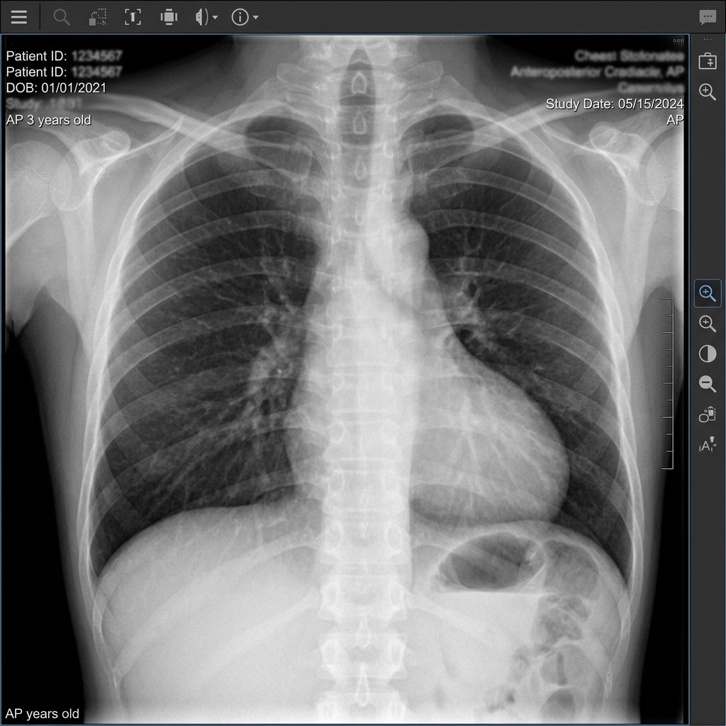
Boot-shaped heart in Tetralogy of Fallot
Posterior-anterior (PA) chest radiograph demonstrating the classic 'Boot-Shaped' cardiac silhouette of Tetralogy of Fallot (TOF), caused by prominent right ventricular hypertrophy lifting the apex and hypoplasia/concavity of the pulmonary artery outflow segment. TOF is the most common cyanotic congenital heart defect (PROVe: Pulmonic stenosis, RVH, Overriding aorta, VSD).
Congenital Cyanotic Heart Defects (The 5 T's)
Clinical Matrix
DefectPathophysiologic AnatomyClassic CXR FindingImmediate Clinical Management
Truncus ArteriosusSingle great vessel arising from ventricles overriding large VSDCardiomegaly, increased pulmonary vascularityDiuretics, surgical repair in first 6 months
Transposition of Great Arteries (TGA)Aorta arises from RV, pulmonary artery from LV; parallel circuits'Egg-on-a-string' cardiac silhouetteIV Prostaglandin E1 (alprostadil) to maintain PDA; Balloon atrial septostomy; Arterial switch
Tricuspid AtresiaAbsent tricuspid valve, hypoplastic RV; ASD and VSD required for survivalDecreased pulmonary vascular markings, left axis deviationIV PGE1 to maintain ductus; Staged Fontan surgical completion
Tetralogy of Fallot (TOF)PROVe: Pulmonic stenosis, RVH, Overriding aorta, VSD'Boot-shaped' heart (concave pulmonary artery segment)Knee-chest position and O2 for 'tet spells' (increases SVR); surgical repair
Total Anomalous Pulm Venous ReturnAll 4 pulmonary veins drain into systemic venous circulation (RA/SVC)'Snowman' appearance or figure-8 on chest radiographSurgical rerouting of pulmonary veins into left atrium
COMLEX Clinical Integration — Cranial Molding & Condylar Compression in the Neonate
COMLEX & OPP Board Integration
During passage through the maternal birth canal, occipital compression against the maternal sacral promontory or pubic bone can cause intraosseous strain of the four parts of the neonatal occiput (squama, two condylar parts, basiocciput). Compression of the occipital condyles against the hypoglossal canal (cranial nerve XII) produces hypoglossal nerve irritation, manifesting clinically as poor suckling, latching difficulties, and tongue thrusting in the newborn. Decompression of the occipital condyles (condylar decompression technique) gently frees the hypoglossal canal, restores tongue motor function, and dramatically improves nursing.

Chapter 1 Quick-Check Self-Assessment

Test your clinical reasoning before moving to the next chapter
3 Vignettes
Board Vignette #1 Single Best Answer
A newborn delivered at 39 weeks is limp and apneic. After drying, warming, and positioning the airway, the heart rate is 70 bpm. What is the single most appropriate next step in management?
Board Vignette #2 Single Best Answer
A 12-hour-old full-term infant becomes acutely cyanotic (SpO2 64%) that fails to improve with 100% FiO2 on hyperoxia test. Echocardiography demonstrates that the aorta arises from the right ventricle and the pulmonary artery arises from the left ventricle. What pharmacologic infusion must be started immediately to sustain life?
Board Vignette #3 Single Best Answer
A term male infant is born to an Rh-negative mother. At 18 hours of life, he appears deeply jaundiced. Total serum bilirubin is 14.8 mg/dL with direct bilirubin 0.4 mg/dL. Direct antiglobulin (Coombs) test is positive. What is the most appropriate initial therapy to prevent kernicterus?
Estimated study time: 14 min
Chapter 2 • Growth, Development & Milestones
12 min TOC

Pediatric Growth, Nutrition & Developmental Milestones

Gross Motor, Fine Motor, Language, and Social-Emotional Milestones (2 Months to 5 Years)

Clinical Overview & Board Focus

Developmental surveillance tracks four core functional streams: Gross Motor, Fine Motor, Language, and Social/Adaptive. Pinpointing developmental delay enables early intervention services. Understanding caloric requirements, failure to thrive (FTT), and childhood immunization schedules is critical for COMAT mastery.

2.1 Chronological Milestone Matrix & Primitive Reflexes

Developmental surveillance tracks four core functional streams: Gross Motor, Fine Motor, Language, and Social/Adaptive. At 2 months: social smile, tracks past midline, lifts head to 45 degrees prone. At 4 months: rolls front-to-back, grasps rattle, laughs, holds head steady. At 6 months: sits unsupported with tripodding, rolls back-to-front, transfers objects hand-to-hand, babbles consonant sounds, stranger anxiety begins. At 9 months: pulls to stand, crawls, pincer grasp (crude 3-finger), plays peek-a-boo, responds to own name, waves 'bye-bye'.

At 12 months (1 year): walks independently or cruises, neat 2-finger pincer grasp, speaks 1–3 specific words ('mama', 'dada'), follows 1-step command with gesture, drinks from a cup. At 18 months: runs, climbs onto chair, builds tower of 3–4 cubes, speaks 10–25 words, uses a spoon. At 2 years (24 months): kicks a ball, walks up and down stairs two feet per step, builds tower of 6 cubes, speaks 2-word phrases ('want milk'), 50% of speech understood by strangers, parallel play.

At 3 years: pedals a tricycle, walks up stairs alternating feet, copies a circle, speaks in 3-word sentences, 75% of speech understood by strangers, dresses with help, group cooperative play. At 4 years: hops on one foot, copies a cross, catches a bounced ball, 100% of speech understood by strangers, tells stories, independent toileting. At 5 years: skips, ties shoes, copies a triangle, prints letters/numbers, counts to 10.

Key Developmental Milestones Master Matrix
Clinical Matrix
AgeGross MotorFine MotorLanguageSocial / Adaptive
2 MonthsLifts head 45° proneHands open, tracks past midlineCoos, vocalizes soundsSocial smile
4 MonthsRolls prone to supineReaches and grasps objectsLaughs, squealsCalms to parent voice
6 MonthsSits unsupportedTransfers hand-to-handBabbles consonant soundsStranger anxiety
9 MonthsPulls to stand, crawlsCrude 3-finger pincer graspUnderstands 'no', babbles mama/dadaPeek-a-boo, object permanence
12 MonthsWalks aloneNeat 2-finger pincer grasp1–3 specific wordsSeparation anxiety, uses cup
2 YearsRuns, kicks ball, stairs 2 feet/stepTower of 6 cubes, turns page2-word telegraphic sentencesParallel play, removes clothes
3 YearsTricycle, stairs alternating feetCopies circle, uses scissors3-word sentences (75% intelligible)Cooperative play, knows age/sex
4 YearsHops on one footCopies cross, draws 4-part personTells stories (100% intelligible)Preferred friends, dressing alone
5 YearsSkips, walks backwardCopies triangle, ties shoesFluent sentences, counts to 10Follows game rules, independent
High-Yield Clinical Takeaway — Red Flag Developmental Signs
High-Yield Clinical Takeaway
Immediate multidisciplinary referral for audiometry, speech evaluation, or neurodevelopmental assessment is warranted for: (1) Absence of babbling or social gestures by 12 months; (2) Inability to sit unsupported by 9 months; (3) Inability to walk independently by 18 months; (4) Failure to speak ≥ 6 meaningful words by 18 months; or (5) ANY loss of previously acquired speech, language, or social skills at any age (concerning for Rett syndrome, autism spectrum disorder, or neurodegenerative regression).

Chapter 2 Quick-Check Self-Assessment

Test your clinical reasoning before moving to the next chapter
3 Vignettes
Board Vignette #1 Single Best Answer
A 2-year-old boy is brought to clinic for a well-child check. His mother is concerned about his development. On exam, he can run, kick a ball, and turn doorknobs. He speaks in 2-word phrases ('more juice'), and his speech is roughly 50% understood by the clinician. He engages in parallel play. How should the clinician counsel the mother regarding his development?
Board Vignette #2 Single Best Answer
A 12-month-old infant is brought to the clinic for a well-child check. The mother is concerned because the infant cannot yet stand alone or walk. Physical examination shows the infant can sit unsupported without falling, pull to a stand while holding a table, transfer a block between hands, use a mature pincer grasp to pick up a cheerio, say 'mama' and 'dada' specifically, and wave 'bye-bye'. Which of the following is the most accurate developmental assessment?
Board Vignette #3 Single Best Answer
During a routine 9-month well-child visit, a healthy infant can sit without support, babble with duplicated syllables ('dada', 'baba'), and demonstrate a crude pincer grasp. When a toy is covered under a blanket, he reaches for it and removes the blanket. What developmental cognitive concept does this demonstrate?
Estimated study time: 12 min
Chapter 3 • Pediatric Infectious Disease & Exanthems
15 min TOC

Pediatric Infectious Diseases, Exanthems & Upper Airway Infections

The Classic Exanthems, Croup vs. Epiglottitis, Meningitis Protocols, and Kawasaki Disease

Clinical Overview & Board Focus

Pediatric fever and rash syndromes require immediate differentiation of life-threatening systemic emergencies (meningococcemia, Kawasaki disease, epiglottitis) from benign self-limiting viral exanthems (roseola, erythema infectiosum).

3.1 The Classic Pediatric Viral & Bacterial Exanthems

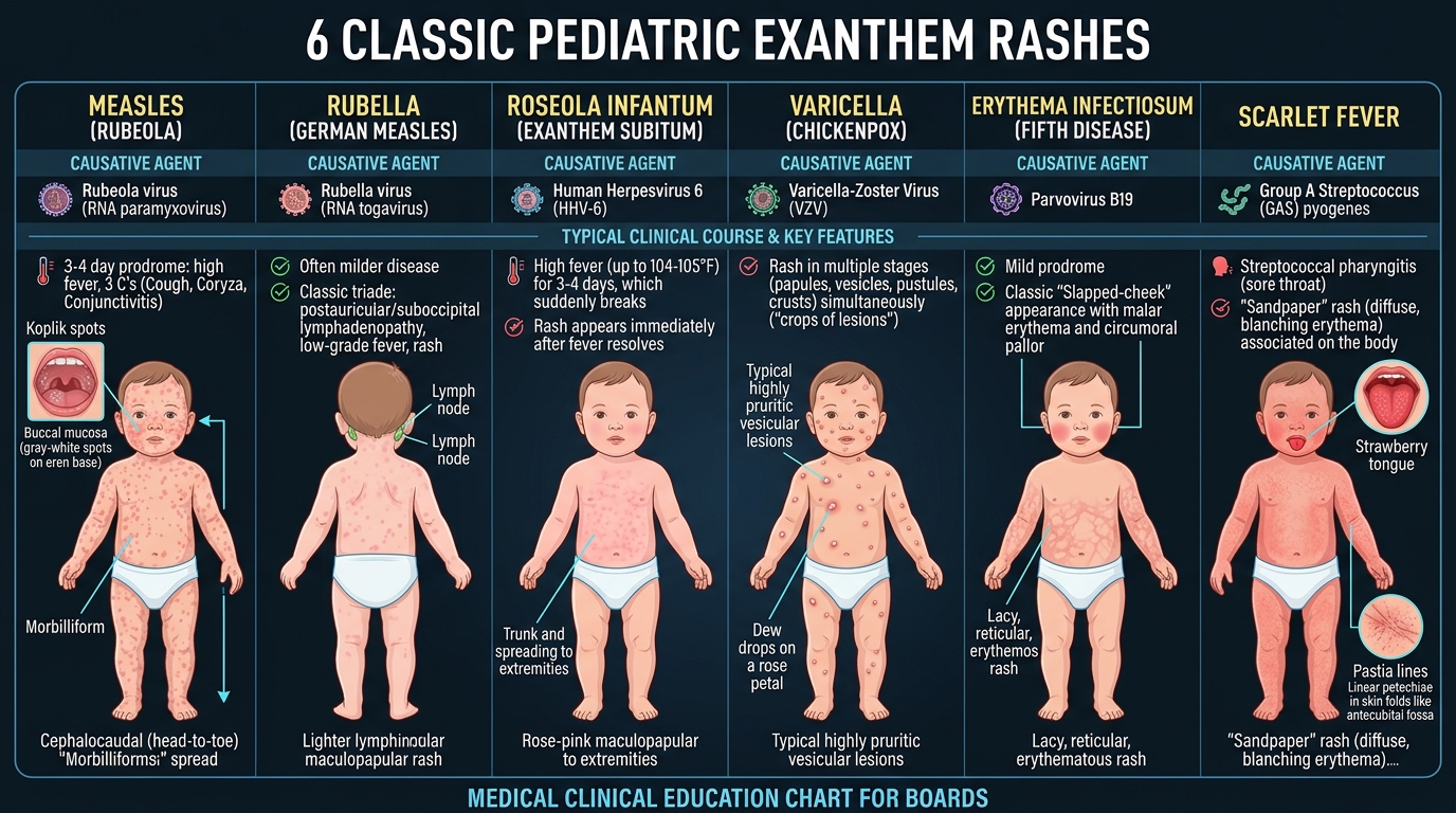
Measles (Rubeola, paramyxovirus) begins with a high prodromal fever accompanied by the classic '3 C's': Cough, Coryza, and Conjunctivitis with photophobia. Pathognomonic Koplik spots (tiny, white-blue grain-of-salt lesions on an erythematous buccal mucosa opposite the lower molars) appear 1–2 days prior to the exanthem. The erythematous maculopapular rash begins at the hairline and face, spreading cephalocaudally down the trunk and extremities, becoming confluent and desquamating. Vitamin A supplementation is mandatory to reduce morbidity and mortality from giant cell pneumonia and blindness. Subacute Sclerosing Panencephalitis (SSPE) is a fatal, late neurodegenerative complication occurring 7–10 years post-infection.

Rubella (German Measles, togavirus) presents with a mild fever, striking tender suboccipital and postauricular lymphadenopathy, and Forchheimer spots (petechiae on the soft palate). A fine pink maculopapular rash spreads rapidly cephalocaudally, clearing from the face as it reaches the trunk. Congenital Rubella Syndrome produces the classic triad: Sensorineural hearing loss, Cataracts, and Cardiac malformations (Patent Ductus Arteriosus [PDA] or peripheral pulmonary stenosis), often accompanied by a 'blueberry muffin' purpuric skin rash from extramedullary hematopoiesis.

Roseola Infantum (Exanthem Subitum, Human Herpesvirus 6 [HHV-6]) classically affects infants aged 6–18 months. It is characterized by 3 to 5 days of exceptionally high fever (> 40°C, often precipitating febrile seizures) in an otherwise well-appearing child. As the fever abruptly defervesces ('breaks'), a faint, rose-pink maculopapular rash appears on the neck and trunk, spreading to the extremities, sparing the face. Erythema Infectiosum (Fifth Disease, Parvovirus B19) presents in school-aged children with bright erythematous facial erythema ('slapped cheek' appearance with circumoral pallor), followed days later by a reticular, lacy, pruritic erythematous rash over the trunk and extremities that waxes and wanes with temperature changes. Parvovirus B19 selectively infects erythrocyte progenitor cells, causing transient aplastic crisis in patients with underlying hemoglobinopathies (sickle cell disease, hereditary spherocytosis) and hydrops fetalis in utero.

6 Classic Pediatric Exanthem Rashes Guide
Comparative clinical atlas of the 6 Classic Pediatric Exanthems: 1) Measles (Rubeola): 3 C's (cough, coryza, conjunctivitis), Koplik spots, cephalocaudal morbilliform rash; 2) Rubella (German Measles): Suboccipital lymphadenopathy; 3) Roseola (HHV-6): High fever resolves, followed immediately by trunk rash; 4) Varicella (VZV): Crops of vesicles at various stages ('dew drops on rose petals'); 5) Erythema Infectiosum (Fifth Disease / Parvovirus B19): 'Slapped-cheek' malar rash followed by lacy reticular body erythema; 6) Scarlet Fever (GAS): Sandpaper rash, Pastia lines, strawberry tongue.
Upper Airway Pediatric Emergencies Comparison
Clinical Matrix
Clinical EntityEtiology / AgeClinical PresentationLateral / AP Neck X-RayDefinitive Management
Croup (Laryngotracheobronchitis)Parainfluenza virus (6 mo – 3 yr)Barking seal-like cough, inspiratory stridor, hoarseness, low fever'Steeple sign' (subglottic tracheal narrowing)Dexamethasone (0.6 mg/kg PO/IM); Nebulized Racemic Epinephrine for stridor at rest
Acute EpiglottitisHaemophilus influenzae type b; Strep spp.High fever, tripod posture, drooling, dysphagia, distress, toxic appearance'Thumbprint sign' (swollen, enlarged epiglottis)Do NOT agitate child; emergent airway in OR by anesthesia/ENT + IV Ceftriaxone + Vancomycin
Bacterial TracheitisStaphylococcus aureus secondary to viral croupHigh fever, toxic appearance, thick purulent secretions, stridor unresponsive to epiSubglottic narrowing with irregular tracheal wall shagginessEndotracheal intubation, tracheal suctioning, IV Vancomycin + Cefepime
Foreign Body AspirationToddler (1–3 yr); peanuts, coins, toysSudden choking event, unilateral wheezing, decreased unilateral breath soundsAir-trapping, hyperinflation, mediastinal shift away on expirationRigid bronchoscopy under general anesthesia
Board Trap — Kawasaki Disease Diagnostic Criteria & Cardiac Aneurysms
High-Yield Board Trap & Alert
Kawasaki Disease (mucocutaneous lymph node syndrome) is a medium-vessel vasculitis. Diagnosis requires Fever lasting ≥ 5 days PLUS at least 4 of the 5 'CRASH' criteria: Conjunctivitis (bilateral, non-exudative, limbic sparing), Rash (polymorphous, non-vesicular), Adenopathy (unilateral cervical > 1.5 cm), Strawberry tongue (oral mucosal erythema, cracked lips), Hand/foot changes (erythema and induration of palms/soles, periungual desquamation). The dreaded complication is coronary artery aneurysms (20–25% if untreated). Treatment is immediate High-Dose IVIG (2 g/kg single infusion) plus High-Dose Aspirin (80–100 mg/kg/day, transitioning to low-dose 3–5 mg/kg/day for 6–8 weeks). Baseline and 6-week echocardiograms are mandatory.

Chapter 3 Quick-Check Self-Assessment

Test your clinical reasoning before moving to the next chapter
3 Vignettes
Board Vignette #1 Single Best Answer
A 3-year-old boy presents with high fever for 6 days, bilateral non-exudative conjunctivitis, a bright red 'strawberry' tongue, erythema and induration of the hands and feet, and a tender 2 cm left cervical lymph node. What is the most critical immediate medical therapy to prevent long-term cardiovascular morbidity?
Board Vignette #2 Single Best Answer
A 4-year-old unvaccinated boy is brought to the clinic with 4 days of high fever (103.5°F), severe rhinorrhea, prominent non-purulent conjunctivitis with photophobia, and a harsh barking cough. Physical exam of the oral cavity reveals tiny white lesions surrounded by an erythematous halo on the buccal mucosa opposite the lower molars. A red maculopapular blanching eruption is beginning behind his ears and at the hairline. Which of the following vitamin deficiencies is associated with severe complications from this infection?
Board Vignette #3 Single Best Answer
A 3-year-old child presents with high fever for 6 days, bilateral non-purulent conjunctival injection, cracked red lips, strawberry tongue, polymorphous trunk rash, cervical lymphadenopathy (1.8 cm), and indurated swelling of hands and feet. What is the primary medical therapy required to prevent coronary artery aneurysms?
Estimated study time: 15 min
Chapter 4 • Pediatric Pulmonology & Allergy
13 min TOC

Pediatric Pulmonology, Bronchiolitis & Cystic Fibrosis

RSV Bronchiolitis, Asthma Action Plans, Cystic Fibrosis Pathogenesis, and Foreign Body Airway Obstruction

Clinical Overview & Board Focus

Respiratory complaints represent the single most common cause of pediatric emergency department visits and hospital admissions. Board mastery demands recognizing the viral pathogenesis of bronchiolitis, step-up outpatient asthma management, and the multi-system manifestations of cystic fibrosis.

4.1 Bronchiolitis, Asthma & Cystic Fibrosis Management

Acute Bronchiolitis is the leading cause of hospitalization in infants < 2 years, predominantly caused by Respiratory Syncytial Virus (RSV). Following a 2–3 day prodrome of rhinorrhea and low-grade fever, infants develop tachypnea, subcostal/intercostal retractions, grunting, nasal flaring, and diffuse bilateral expiratory wheezing with fine crackles. Diagnosis is purely clinical; viral PCR swabs, chest X-rays, and blood work are NOT routinely recommended. AAP clinical practice guidelines emphasize supportive care: nasal suctioning, hydration, and supplemental oxygen targeting SpO2 ≥ 90%. Routine administration of bronchodilators (albuterol), epinephrine, and systemic corticosteroids is NOT recommended due to lack of clinical efficacy. Palivizumab (monoclonal antibody against RSV F-protein) provides monthly passive immunoprophylaxis for high-risk preterm infants (< 29 weeks gestation) or those with congenital heart disease or chronic lung disease.

Cystic Fibrosis (CF) is an autosomal recessive disorder caused by mutations in the CFTR gene (most commonly Delta-F508 on chromosome 7), resulting in defective cAMP-regulated chloride and bicarbonate conductance across apical epithelial membranes. In the lungs, thick dehydrated secretions impair mucociliary clearance, resulting in chronic colonization with Staphylococcus aureus in infancy and Pseudomonas aeruginosa in adolescents and adults, leading to bronchiectasis and respiratory failure. In the gastrointestinal tract, exocrine pancreatic insufficiency causes fat malabsorption (steatorrhea, deficiency of fat-soluble vitamins A, D, E, K), failure to thrive, and meconium ileus (inspissated meconium obstructing the terminal ileum, demonstrated as microcolon on contrast enema) in up to 20% of newborns.

Definitive diagnosis of CF is made via Quantitative Pilocarpine Iontophoresis (Sweat Chloride Test): sweat chloride concentration ≥ 60 mmol/L on ≥ 2 occasions confirms the diagnosis. Treatment requires a multidisciplinary approach: airway clearance chest physiotherapy, inhaled dornase alfa (DNase) to reduce mucus viscosity, inhaled hypertonic saline, pancreatic enzyme replacement therapy (PERT) administered with all meals, fat-soluble vitamin supplementation, and targeted CFTR modulators (e.g., Elexacaftor-Tezacaftor-Ivacaftor for patients with at least one Delta-F508 allele).

COMLEX Clinical Integration — Rib Raising & Diaphragmatic Release in Pediatric Bronchospasm
COMLEX & OPP Board Integration
In pediatric asthma and reactive airway diseases, hyperinflation and coughing cause marked facilitation of sympathetic ganglia from T1 to T6. Rib raising along the thoracic chain normalizes excessive sympathetic outflow to the tracheobronchial tree, reducing bronchial mucosal secretions and hyperreactivity. Diaphragmatic doming releases the crura (anchored at L1–L3) and improves lymphatic drainage through the cisterna chyli into the left venous angle. Suboccipital release targets vagal parasympathetics (cranial nerve X) at the jugular foramen to restore bronchomotor equilibrium.

Chapter 4 Quick-Check Self-Assessment

Test your clinical reasoning before moving to the next chapter
3 Vignettes
Board Vignette #1 Single Best Answer
A 9-month-old infant is diagnosed with Cystic Fibrosis following a positive sweat chloride test (84 mmol/L). What is the premier bacterial pathogen responsible for recurrent pulmonary infections and pneumonia during the first decade of life in this child?
Board Vignette #2 Single Best Answer
A 3-year-old unvaccinated boy is brought to the emergency department with acute high fever (104.2°F), difficulty swallowing, muffled voice, and inspiratory stridor. He is sitting upright, leaning forward with his neck extended and mouth open, and saliva is drooling onto his shirt. What is the most appropriate next step in management?
Board Vignette #3 Single Best Answer
A 2-year-old boy presents with sudden barking cough, inspiratory stridor at rest, and mild intercostal retractions. AP neck radiograph demonstrates symmetric subglottic narrowing ('steeple sign'). In addition to single-dose oral dexamethasone, what is the most appropriate immediate therapy for stridor at rest?
Estimated study time: 13 min
Chapter 5 • Pediatric Gastroenterology & Nutrition
14 min TOC

Pediatric Gastroenterology, Intestinal Obstruction & Malabsorption

Hypertrophic Pyloric Stenosis, Intussusception, Malrotation with Midgut Volvulus, Hirschsprung Disease, and Celiac Disease

Clinical Overview & Board Focus

Pediatric gastrointestinal emergencies center on distinguishing non-bilious from bilious vomiting, identifying congenital anatomical obstructions, and evaluating neonatal abdominal distention. Board exams test physical examination palpation, diagnostic ultrasound features, and contrast fluoroscopy findings.

5.1 Mechanical Bowel Obstruction in Infants & Children

Hypertrophic Pyloric Stenosis results from hypertrophy and hyperplasia of the circular pyloric sphincter muscle, classically presenting in first-born males at 3 to 6 weeks of life with progressive, forceful, projectile non-bilious vomiting immediately after feedings. The infant remains ravenously hungry ('hungry vomiter'). Physical examination reveals an olive-shaped, mobile mass palpated in the right upper quadrant or epigastrium. Transverse abdominal ultrasonography is diagnostic, demonstrating pyloric muscle thickness ≥ 3 mm and channel length ≥ 14 mm. Persistent vomiting of gastric hydrochloric acid produces the classic serum metabolic profile: Hypochloremic, Hypokalemic Metabolic Alkalosis. Surgical pyloromyotomy (Ramstedt procedure) is curative, but must NEVER be performed until intravenous fluid rehydration completely normalizes serum chloride, potassium, and acid-base status.

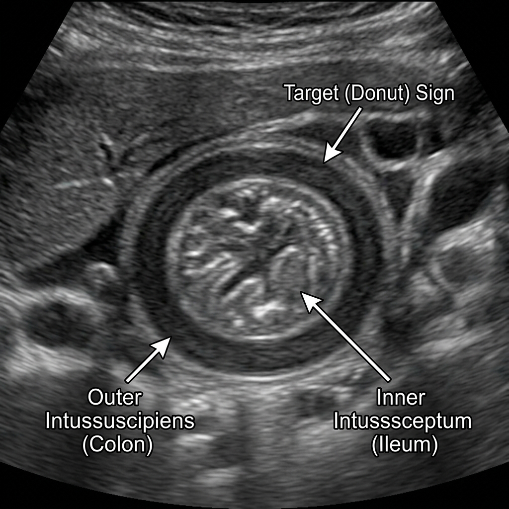
Intussusception involves telescoping of a proximal bowel segment into an adjacent distal segment (most commonly ileocolic), typically occurring between 6 months and 3 years of age. It presents with sudden, episodic, severe colicky abdominal pain during which the infant draws their knees up to their chest and cries inconsolably, alternating with periods of lethargy. A sausage-shaped abdominal mass is palpated in the right upper quadrant with emptiness in the right lower quadrant (Dance sign). Passage of 'currant jelly stools' (blood and sloughed mucus) is a late sign indicating mucosal ischemia. Abdominal ultrasound is the imaging modality of choice, demonstrating a 'target' or 'doughnut' sign in transverse view. In the absence of peritonitis or bowel perforation, pneumatic (air-contrast) or hydrostatic (barium/saline) enema is both diagnostic and therapeutic (reducing the intussusception in > 85% of cases). In children > 3 years, look for a pathologic lead point (Meckel diverticulum, Henoch-Schönlein purpura hematoma, or Burkitt lymphoma).

Malrotation with Midgut Volvulus occurs due to incomplete 270-degree counterclockwise embryologic rotation of the midgut around the superior mesenteric artery, leaving a narrow mesenteric base anchored by peritoneal Ladd bands crossing the duodenum. Volvulus can present catastrophically at any time (60% in first month of life) with sudden-onset acute bilious (green-yellow) vomiting and abdominal distention in a previously thriving infant. Upper GI fluoroscopy series is the gold standard imaging modality, demonstrating duodenal 'corkscrew' appearance and failure of the duodenojejunal junction (ligament of Treitz) to cross the left midline. Midgut volvulus is a surgical emergency; immediate exploratory laparotomy with Ladd procedure (counterclockwise untwisting, division of Ladd bands, widening of the mesentery, and appendectomy) is life-saving.

Intussusception Target Sign Ultrasound
Abdominal ultrasound demonstrating a transverse concentric ring appearance—the pathognomonic 'Target' or 'Donut' sign of Intussusception. The telescoping of the ileum into the cecum (ileocecal) causes venous congestion, colicky abdominal pain with legs drawn to chest, and 'currant jelly' stools. Diagnostic and therapeutic intervention of choice: Air or pneumatic contrast enema under fluoroscopic/ultrasound guidance.
Pediatric Bowel Obstruction Differential Matrix
Clinical Matrix
EntityAge of OnsetVomitus CharacterClassic Physical / Lab FindingDiagnostic Modality / Treatment
Pyloric Stenosis3–6 WeeksNon-bilious, projectile'Olive' mass in RUQ; hypochloremic hypokalemic alkalosisUltrasound (pyloric thickness ≥ 3 mm); IV hydration → Pyloromyotomy
Intussusception6–36 MonthsBilious or non-biliousSausage-shaped RUQ mass; 'currant jelly' stool; intermittent colicUltrasound ('target sign'); Air-contrast enema reduction
Midgut VolvulusFirst month of lifeBilious (dark green)Abdominal distention, rapid progression to septic peritonitisUpper GI series ('corkscrew' duodenum); Emergent Ladd procedure
Hirschsprung DiseaseFirst 48 hr of lifeBilious vomitingFailure to pass meconium in 48 hr; 'blast sign' on rectal examContrast enema (transition zone); Suction Rectal Biopsy (absent ganglion cells)
Duodenal AtresiaFirst 24–48 hrBilious vomitingAssociated with Down syndrome (Trisomy 21); maternal polyhydramniosAbdominal X-ray ('double bubble' sign); Duodenoduodenostomy
Board Trap — The 'Blast Sign' in Hirschsprung Disease
High-Yield Board Trap & Alert
Hirschsprung disease (congenital aganglionic megacolon) results from failure of neural crest cells to migrate cranio-caudally into the myenteric (Auerbach) and submucosal (Meissner) plexuses of the distal rectum and sigmoid. Digital rectal examination reveals a tight anal sphincter with an empty rectal vault; upon withdrawal of the finger, there is an explosive expulsion of gas and liquid stool ('blast sign' / squirt sign). The gold standard for definitive diagnosis is Suction Rectal Biopsy showing absent ganglion cells and hypertrophied non-myelinated nerve fibers with positive acetylcholinesterase staining.

Chapter 5 Quick-Check Self-Assessment

Test your clinical reasoning before moving to the next chapter

3 Vignettes
Board Vignette #1 Single Best Answer
A 4-week-old first-born male infant presents with forceful projectile non-bilious vomiting after every feeding for 4 days. Serum electrolytes demonstrate Na 134 mEq/L, K 2.8 mEq/L, Cl 86 mEq/L, and HCO3 34 mEq/L. Abdominal ultrasound confirms pyloric muscle thickness of 4.5 mm. What must be completed prior to proceeding to surgical pyloromyotomy?
Board Vignette #2 Single Best Answer
A 4-week-old first-born male infant presents with forceful, non-bilious projectile vomiting occurring immediately after every feeding for the past 5 days. He is voraciously hungry immediately after vomiting. Physical examination reveals a small, firm, mobile 1.5-cm olive-shaped mass in the right upper quadrant. Serum chemistry reveals sodium 134 mEq/L, potassium 3.0 mEq/L, chloride 86 mEq/L, and bicarbonate 32 mEq/L. Which of the following is the most appropriate management prior to definitive surgical pyloromyotomy?
Board Vignette #3 Single Best Answer
A 3-week-old first-born male infant presents with forceful non-bilious projectile vomiting immediately after every feed. He is vigorously hungry. Physical exam reveals a firm, non-tender 2-cm olive-shaped mass in the right epigastrium. Serum chemistry shows sodium 134, potassium 2.9, chloride 84, and bicarbonate 34 mEq/L. What is the most critical initial management step prior to surgical pyloromyotomy?
Estimated study time: 14 min
Chapter 6 • Pediatric Nephrology, Fluids & Urology
13 min TOC

Pediatric Nephrology, Hematuria & Fluid Balance

Minimal Change Disease, Post-Streptococcal Glomerulonephritis, Hemolytic Uremic Syndrome, and Vesicoureteral Reflux

Clinical Overview & Board Focus

Pediatric renal pathology partitions into nephrotic syndromes (massive proteinuria, hypoalbuminemia, edema, hyperlipidemia) and nephritic syndromes (hematuria, RBC casts, hypertension, oliguria). Board examinations heavily test fluid maintenance calculations (Holliday-Segar method), post-infectious glomerulonephritis, and microangiopathic hemolytic anemia in HUS.

6.1 Nephrotic Syndrome, Nephritic Glomerulopathies & HUS

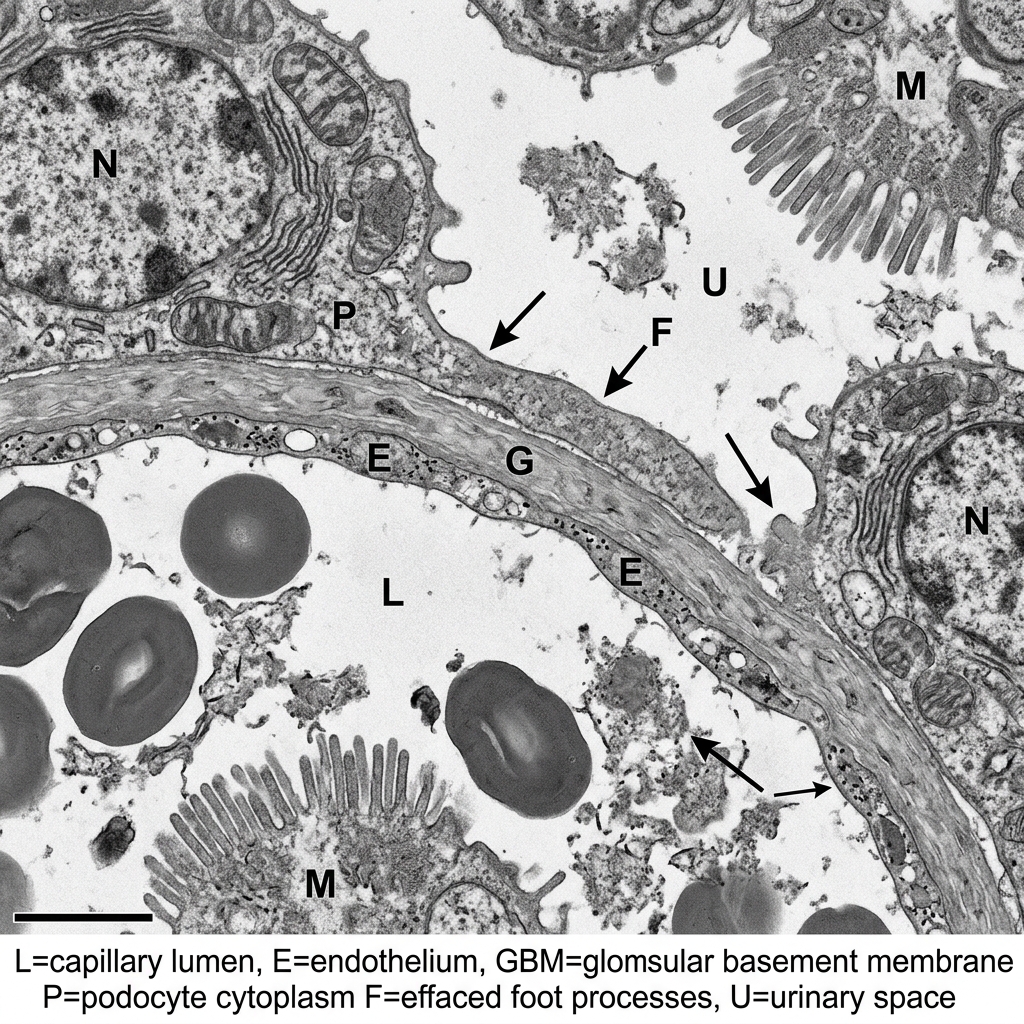
Minimal Change Disease (MCD) accounts for > 85% of nephrotic syndrome cases in children aged 2 to 6 years. It is characterized by T-cell mediated cytokine release that causes loss of negatively charged heparan sulfate proteoglycans in the glomerular basement membrane, resulting in selective albuminuria. Children present with periorbital edema that progresses to dependent peripheral edema, ascites, and pleural effusions. Laboratory evaluation confirms the nephrotic tetrad: massive proteinuria (spot urine protein-to-creatinine ratio > 2.0 or 24-hr urine protein > 50 mg/kg/day), profound hypoalbuminemia (< 2.5 g/dL), generalized edema, and secondary hypercholesterolemia. Renal biopsy is NOT indicated in typical presentations; light microscopy is normal, and electron microscopy reveals diffuse effacement of podocyte foot processes. First-line therapy is oral Corticosteroids (Prednisone 2 mg/kg/day × 6 weeks), resulting in complete remission in > 90% of children. Patients are at high risk for encapsulated bacterial peritonitis (Streptococcus pneumoniae) due to urinary loss of immunoglobulins and complement factors, and thromboembolism due to antithrombin III urinary wasting.

Post-Streptococcal Glomerulonephritis (PSGN) is an immune-complex mediated nephritic syndrome occurring 1 to 3 weeks after Group A beta-hemolytic Streptococcal (Streptococcus pyogenes) pharyngitis or 3 to 6 weeks after streptococcal impetigo. Children present with the acute nephritic triad: macroscopic hematuria ('tea-colored' or 'cola-colored' urine with dysmorphic RBCs and RBC casts), periorbital edema, and hypertension with mild oliguria. Laboratory workup reveals low serum complement C3 (which normalizes within 6–8 weeks) and elevated streptococcal antibody titers (anti-streptolysin O [ASO] following pharyngitis; anti-DNase B following impetigo). Light microscopy demonstrates enlarged, hypercellular glomeruli with neutrophil infiltration; immunofluorescence shows granular 'lumpy-bumpy' deposits of IgG and C3; electron microscopy reveals subepithelial 'humps'. Treatment is supportive (fluid and sodium restriction, loop diuretics for fluid overload and hypertension); prognosis in children is excellent (> 95% full recovery).

Hemolytic Uremic Syndrome (HUS) is the leading cause of acute pediatric renal failure, triggered in > 90% of cases by Shiga toxin-producing Escherichia coli (STEC, serotype O157:H7) following ingestion of undercooked ground beef or unpasteurized juice, or Shigella dysenteriae. Following a 3–5 day prodrome of bloody diarrhea and abdominal cramping, the child develops the classic HUS triad: (1) Microangiopathic Hemolytic Anemia (schistocytes, helmet cells on peripheral blood smear, elevated LDH, indirect hyperbilirubinemia, low haptoglobin, negative direct Coombs test); (2) Thrombocytopenia (platelets < 50,000 without petechiae or deep bleeding); and (3) Acute Renal Failure (oliguria, azotemia, hematuria, proteinuria). Treatment is strictly supportive (cautious isotonic fluid hydration, packed RBC transfusions as needed, and hemodialysis in up to 50%). Antibiotics and antimotility agents (loperamide) are STRICTLY CONTRAINDICATED because they increase Shiga toxin release from lysed bacteria, worsening renal thrombotic microangiopathy.

Minimal Change Disease Podocyte Foot Process Effacement TEM
Transmission Electron Microscopy (TEM) of the glomerular filtration barrier in Minimal Change Disease (MCD) demonstrating diffuse, extensive effacement (flattening) of the visceral epithelial podocyte foot processes with loss of intervening filtration slits. Light microscopy appears completely normal. MCD is the most common cause of pediatric nephrotic syndrome (>85%), highly responsive to oral corticosteroid therapy.
Board Trap — Antibiotics in E. coli O157:H7 Enteritis
High-Yield Board Trap & Alert
A 4-year-old child presents with 4 days of severe bloody diarrhea and cramping after visiting a petting zoo. Stool PCR identifies Shiga toxin-producing E. coli (STEC). Prescribing fluoroquinolones, beta-lactams, or macrolides triggers bacterial stress and lysis, precipitating a massive surge in systemic Shiga toxin absorption and dramatically increasing the risk of developing full-blown Hemolytic Uremic Syndrome and renal failure. Withhold antibiotics; provide aggressive supportive isotonic IV hydration.

Chapter 6 Quick-Check Self-Assessment

Test your clinical reasoning before moving to the next chapter

3 Vignettes
Board Vignette #1 Single Best Answer
A 4-year-old boy presents with progressive periorbital swelling and swollen ankles. Urinalysis reveals 4+ protein and 0 RBCs. Spot urine protein-to-creatinine ratio is 4.5. Serum albumin is 2.1 g/dL and total cholesterol is 320 mg/dL. What is the most appropriate next step in management?
Board Vignette #2 Single Best Answer
A 7-year-old boy is brought to the clinic because of dark, 'tea-colored' urine and puffy eyes for 2 days. Two weeks ago, he was treated for impetigo on his face. On examination, blood pressure is 132/84 mmHg and there is 1+ pretibial and periorbital edema. Urinalysis shows 3+ blood, 1+ protein, and dysmorphic red blood cells with red blood cell casts. Serum complement C3 is markedly decreased. What is the characteristic finding on electron microscopy of a renal biopsy?
Board Vignette #3 Single Best Answer
A 5-year-old boy presents with palpable purpura on the lower extremities and buttocks, colicky abdominal pain, and swelling of both ankles 10 days following an upper respiratory infection. Platelet count and coagulation profile (PT/INR) are completely normal. Urinalysis reveals 2+ proteinuria and microhematuria. What is the underlying pathophysiology?
Estimated study time: 13 min
Chapter 7 • Pediatric Musculoskeletal & Orthopedics
14 min TOC

Pediatric Orthopedics & The Limping Child

Developmental Dysplasia of the Hip, Slipped Capital Femoral Epiphysis, Legg-Calvé-Perthes, and Septic Arthritis vs. Transient Synovitis

Clinical Overview & Board Focus

Evaluating the limping child requires age-stratified differential diagnosis and immediate exclusion of septic arthritis. Board exams prioritize clinical hip maneuvers in newborns, Kocher criteria for septic hip, and the radiographic hallmark of SCFE.

7.1 The Limping Child & Pediatric Hip Pathology Matrix

Developmental Dysplasia of the Hip (DDH) involves abnormal development of the femoral head and acetabulum, ranging from subluxation to complete dislocation. Risk factors include breech presentation, female sex, and family history. Screening physical examination maneuvers performed in infancy are: Barlow maneuver (flex and adduct the hip while applying posterior force; attempts to 'dislocate' the unstable hip) and Ortolani maneuver (flex and abduct the hip while applying anterior force on greater trochanter; 'relocates' the dislocated hip with a palpable 'clunk'). After 3 months of age, Galeazzi test (unequal knee heights with hips and knees flexed) and asymmetric thigh skin folds become prominent. Ultrasound of the hip is the imaging modality of choice before 4–6 months (prior to femoral head ossification); pelvic X-rays are used after 6 months. First-line management is a Pavlik harness (splint maintaining hip flexion and abduction) for infants < 6 months.

Transient Synovitis (toxic synovitis) is a benign, self-limiting reactive inflammation of the hip synovial membrane, classically occurring in boys aged 3 to 8 years following a viral upper respiratory infection. The child presents with a limp or refusal to bear weight, mild hip pain, and is afebrile or low-grade. Ultrasound shows a small joint effusion. In sharp contrast, Septic Arthritis of the hip is an orthopedic emergency requiring immediate surgical drainage to prevent femoral head necrosis. Differentiating between the two relies on the Kocher Criteria: (1) Fever > 38.5°C (101.3°F); (2) Inability to bear weight; (3) Erythrocyte sedimentation rate (ESR) > 40 mm/hr; and (4) Serum white blood cell (WBC) count > 12,000/µL (CRP > 2.0 mg/dL added as 5th criterion). The presence of 4 of 4 Kocher criteria carries a > 99% probability of septic arthritis, mandating immediate ultrasound-guided arthrocentesis (synovial fluid WBC > 50,000/µL with > 75% PMNs confirms septic arthritis) and emergent operative arthrotomy.

Legg-Calvé-Perthes Disease is idiopathic avascular necrosis (osteonecrosis) of the femoral head, classically affecting active boys aged 4 to 10 years. It presents with insidious-onset, painless or mildly painful limp exacerbated by activity, with loss of hip internal rotation and abduction. Early pelvis AP and frog-leg lateral radiographs may be normal or show subtle crescent sign; late stages demonstrate femoral head flattening, fragmentation, and sclerosis. Slipped Capital Femoral Epiphysis (SCFE) is the posteromedial displacement of the femoral head through the growth plate (physis), classically occurring in obese adolescent males (aged 10–16 years) during peak pubertal growth spurt. Patients present with dull groin, thigh, or isolated knee pain (referred pain via the obturator nerve) with an antalgic limp. Physical exam reveals obligatory external rotation of the hip during passive hip flexion. Frog-leg lateral pelvis X-ray confirms the slip (Klein's line fails to intersect the lateral aspect of the femoral epiphysis). SCFE is an orthopedic emergency; immediate non-weight-bearing crutches and emergent in situ percutaneous single cannulated screw fixation are required to prevent avascular necrosis and chondrolysis.

Pediatric Hip Orthopedic Examination
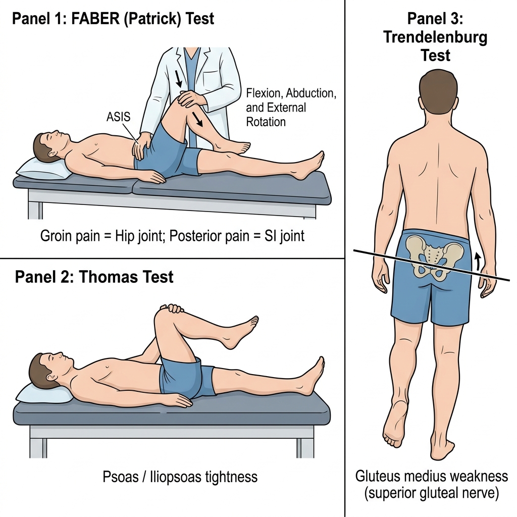
Pediatric orthopedic hip assessment for Developmental Dysplasia of the Hip (DDH): Barlow test (adduction and posterior pressure to dislocate the femoral head) and Ortolani test (abduction and anterior elevation to relocate the hip with a palpable 'clunk'). In infants < 6 months with abnormal exam or risk factors (breech birth, female), ultrasound of the hip is diagnostic; treatment is the Pavlik harness.
Pediatric Hip Pathology by Age Group
Clinical Matrix
DisorderTypical Age & DemographicsClinical Presentation & ExamKey Imaging HallmarkFirst-Line Management
DDHNewborn to 6 months; females, breechPositive Ortolani / Barlow test; Galeazzi signHip Ultrasound (< 4–6 mo); Pelvic X-ray (> 6 mo)Pavlik harness in flexion and abduction
Transient Synovitis3–8 Years; boys; post-viral URIMild limp, afebrile/low-grade, hip held in flexion/external rotationUltrasound shows small effusion; normal labsBowel rest, NSAIDs, weight-bearing as tolerated; self-limiting
Septic ArthritisAny age (peak < 3 yr)Refusal to bear weight, high fever, extreme pain on passive motionSynovial WBC > 50,000/µL with > 75% neutrophilsEmergent operative joint drainage/washout + IV Ceftriaxone/Vancomycin
Legg-Calvé-Perthes4–10 Years; active boysInsidious painless limp or groin pain; decreased internal rotationX-ray shows femoral head collapse, sclerosis, fragmentationProtected weight-bearing, physical therapy; osteotomy if severe
SCFE10–16 Years; obese adolescent malesDull groin/knee pain; external rotation on passive flexionFrog-leg lateral X-ray: Klein line abnormal; 'ice cream falling off cone'Immediate non-weight bearing + Emergent surgical in situ screw fixation
Board Trap — Knee Pain Heralding Hip Pathology in Adolescents
High-Yield Board Trap & Alert
An obese 12-year-old boy presents to clinic complaining solely of dull right knee pain for 3 weeks, without prior trauma. Knee exam is entirely normal without effusion, ligamentous laxity, or joint-line tenderness. Because sensory branches of the obturator and femoral nerves innervate both the hip joint capsule and medial knee, hip pathology frequently presents exclusively as referred knee pain! Any child with unexplained knee or distal thigh pain must undergo a dedicated hip exam (checking passive internal rotation and flexion) and bilateral hip radiographs to rule out Slipped Capital Femoral Epiphysis.

Chapter 7 Quick-Check Self-Assessment

Test your clinical reasoning before moving to the next chapter

3 Vignettes
Board Vignette #1 Single Best Answer
A 13-year-old obese boy (BMI 33 kg/m²) presents with dull left knee and thigh pain for 4 weeks. He has an antalgic gait. Knee examination is normal. Passive flexion of the left hip causes obligatory external rotation. Bilateral frog-leg lateral pelvic radiographs demonstrate posterior displacement of the left femoral epiphysis. What is the mandatory next step?
Board Vignette #2 Single Best Answer
A 13-year-old obese male presents with a 3-week history of a dull, aching pain in his left knee and an antalgic limp. He denies prior knee trauma. Physical examination of the left knee reveals no swelling, effusion, or joint line tenderness. However, examination of the left hip reveals significant limitation of internal rotation and abduction, with obligate external rotation upon passive hip flexion. What is the most appropriate next step in management?
Board Vignette #3 Single Best Answer
A 13-year-old obese male presents with a 3-week history of a dull, achy left hip and knee pain and an antalgic limp. On physical examination, the left lower extremity is held in external rotation, and passive internal rotation of the hip is severely limited and painful. What is the definitive treatment?
Estimated study time: 14 min