Skip to content
NBOME OPP / OMM COMAT & Level 2-CE NBME OPP / OMM Shelf Exam & Step 2 CK

OPP / OMM COMAT Command Center OPP / OMM Shelf Exam Command Center

Comprehensive NBOME COMAT OPP & Level 1/Level 2-CE osteopathic board mastery center. Synthesizes 670+ board-curated OMM questions across sacral/innominate biomechanics, rib mechanics, autonomics, Chapman reflexes, and cranial osteopathy.

70 Cards Extracted 3 High-Yield Protocols 4 Osteopathic Rules 4 High-Yield Pearls
Section 01 • Osteopathic Principles

"Don't Miss" COMAT Osteopathic Pearls

High-frequency somatic dysfunctions, viscerosomatics, autonomic reflexes, and treatment rules

Tested heavily on NBOME Shelf

The Golden L5 / Sacrum Biomechanical Rules

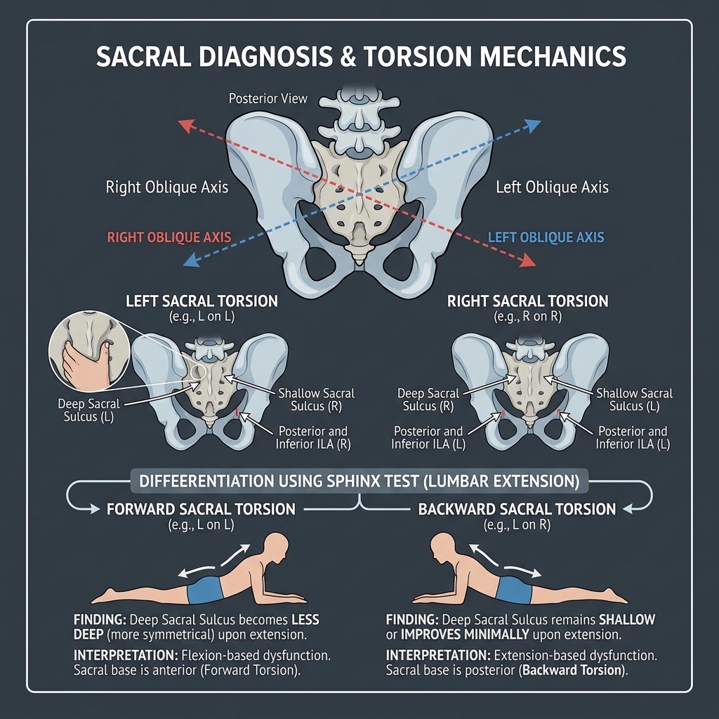
1. L5 Sidebending and the Sacral Oblique Axis are on the SAME side. 2. L5 Rotation and Sacral Rotation are in OPPOSITE directions. 3. Positive seated flexion test occurs on the side opposite the oblique axis (side of axis = negative seated flexion test). Example: L5 rotated right, sidebent left -> indicates a Left Oblique Axis; with sacrum rotated left (opposite L5 rotation), this yields a Left-on-Left forward sacral torsion.

Inhalation vs. Exhalation Rib Dysfunctions: Key Rib Rules

Inhalation Somatic Dysfunction: Ribs are locked in inhalation (stop moving first in exhalation). The KEY rib to treat first is the LOWEST rib in the group ('B-I-L-L': Bottom Inhalation). Exhalation Somatic Dysfunction: Ribs are locked in exhalation (stop moving first in inhalation). The KEY rib to treat first is the TOP/HIGHEST rib in the group ('B-I-L-L': Top Exhalation). Rib 1 uses Anterior/Middle Scalenes; Rib 2 uses Posterior Scalene; Ribs 3–5 use Pectoralis Minor; Ribs 6–9 use Serratus Anterior; Ribs 10–11 use Latissimus Dorsi; Rib 12 uses Quadratus Lumborum.

Autonomic Nervous System Viscerosomatic Level Map

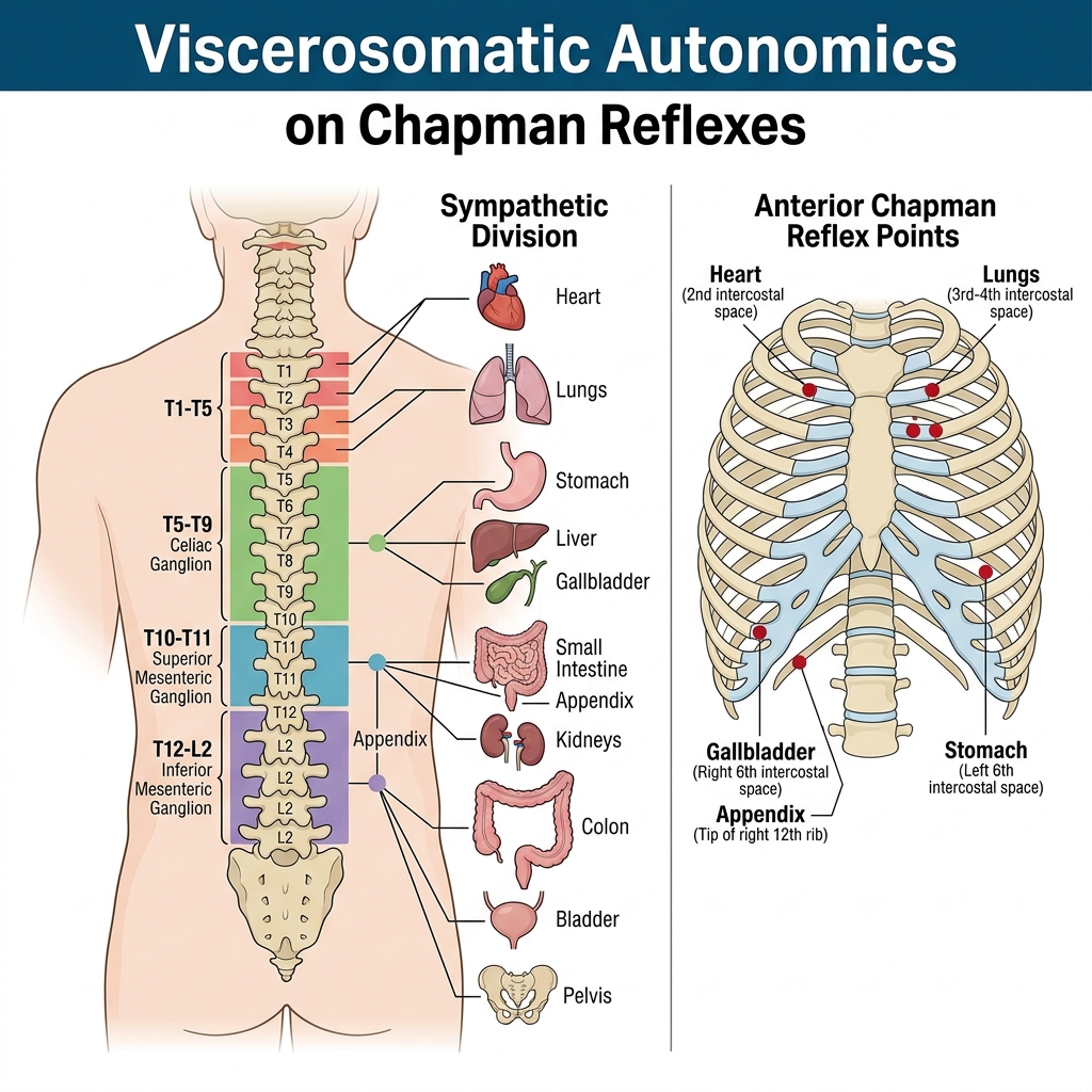
Sympathetics: T1–T5 = Head, Neck, Heart, Lungs; T5–T9 = Upper GI / Foregut (Celiac Ganglion: stomach, liver, gallbladder, spleen, pancreas, proximal duodenum); T10–T11 = Midgut / Gonads / Kidneys / Upper Ureters (SMG: distal duodenum, jejunum, ileum, ascending colon, proximal 2/3 transverse colon); T12–L2 = Hindgut / Pelvic Organs / Lower Ureters / Bladder / Uterus / Prostate (IMG: distal 1/3 transverse colon, descending/sigmoid colon, rectum). Parasympathetics: Vagus (CN X) innervates occiput to proximal 2/3 transverse colon and kidneys; Pelvic Splanchnics (S2–S4) innervate distal 1/3 transverse colon to rectum and pelvic viscera.

Absolute vs. Relative Contraindications to HVLA

Absolute Contraindications: Suspected or confirmed bony fracture, acute dislocation, cervical spine ligamentous instability (Down syndrome, rheumatoid arthritis with transverse ligament of atlas laxity), severe acute osteomyelitis, bone malignancy/metastasis, acute joint infection, patient refusal. Relative Contraindications: Acute disc herniation with radiculopathy, moderate-to-severe osteoporosis, patients on therapeutic anticoagulation, mild hypermobility.

Section 01 • Clinical Foundations

"Don't Miss" OPP / OMM Clinical Pearls & Shelf Traps

High-frequency diagnostic pitfalls, gold-standard criteria, and next-best-step clinical rules

Tested heavily on NBME Shelf & Step 2 CK

Low Back Pain Differential & Neurologic Root Levels

L4 Root: Motor = Quadriceps / Tibialis anterior (knee extension, heel walk, ankle dorsiflexion); Reflex = Patellar tendon; Sensory = Medial leg and medial foot. L5 Root: Motor = Extensor hallucis longus (great toe dorsiflexion, heel walk); Reflex = None; Sensory = Dorsum of foot and first web space. S1 Root: Motor = Gastrocnemius / Soleus / Peroneus longus (toe walk, ankle plantarflexion); Reflex = Achilles tendon; Sensory = Lateral foot and sole. Red flag cauda equina: emergent decompression within 48h.

Shoulder Impingement & Rotator Cuff Tear Examination

Supraspinatus: Empty Can (Jobe) test; Infraspinatus & Teres Minor: Resisted external rotation (Hornblower sign); Subscapularis: Bear-Hug, Belly-Press, or Lift-Off test; Subacromial Impingement: Neer sign (passive forward flexion with pronation) and Hawkins-Kennedy test (internal rotation at 90° flexion). Long head of Biceps: Speed test (resisted flexion with supination) and Yergason test (resisted supination).

Knee Ligamentous & Meniscal Tear Diagnostics

Anterior Cruciate Ligament (ACL): Lachman test (most sensitive, 30° flexion) and Anterior Drawer; Posterior Cruciate Ligament (PCL): Posterior Drawer and Sag sign (dashboard injury); Medial Collateral Ligament (MCL): Valgus stress at 30°; Lateral Collateral Ligament (LCL): Varus stress at 30°; Meniscal Tears: McMurray test (external rotation + extension for medial meniscus; internal rotation + extension for lateral meniscus) and Thessaly test (standing weight-bearing twist).

Hip & Pelvic Physical Examination Maneuvers

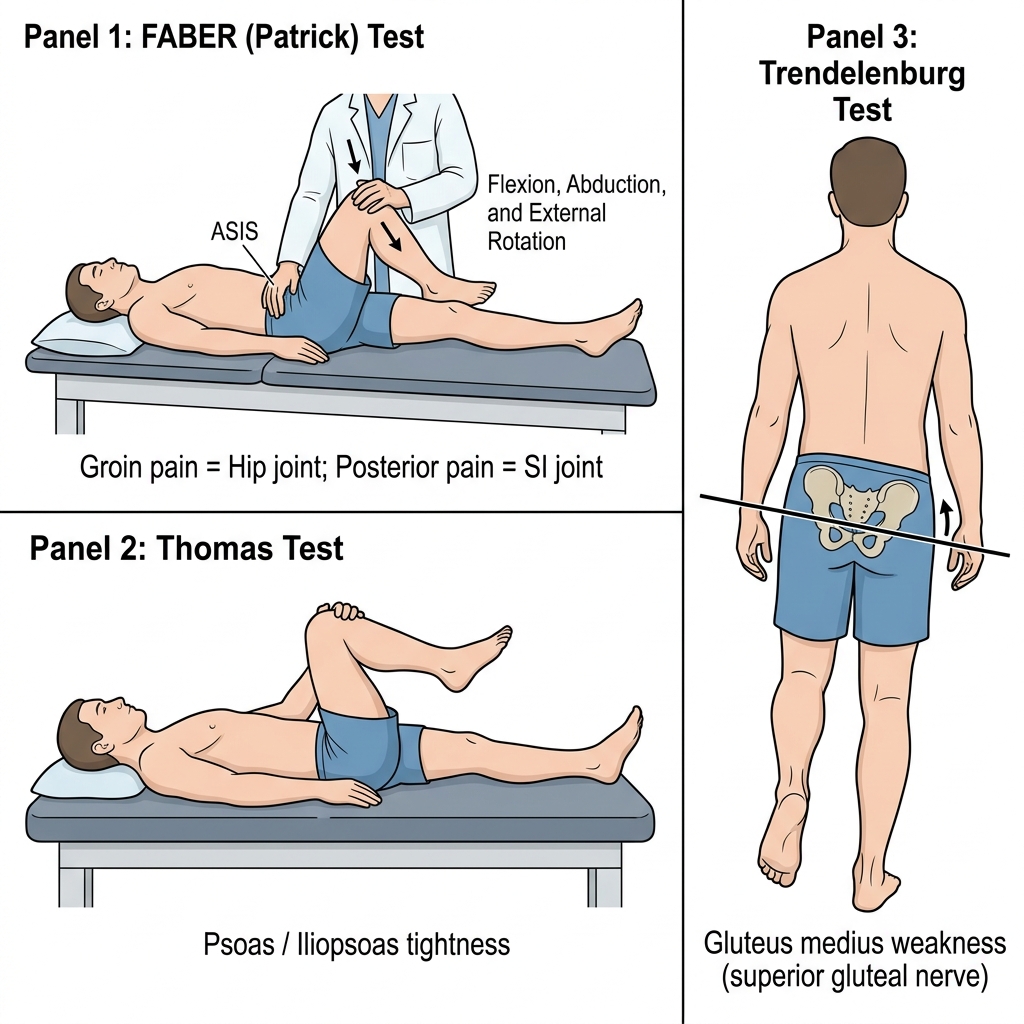
FABER (Patrick) test: Flexion, Abduction, External Rotation (pain anteriorly indicates intra-articular hip pathology/osteoarthritis; pain posteriorly indicates sacroiliac joint pathology). FADIR test: Flexion, Adduction, Internal Rotation (sensitive for femoroacetabular impingement [FAI] or labral tear). Trendelenburg sign: Dropping of contralateral pelvis on single-leg stance due to ipsilateral gluteus medius weakness (superior gluteal nerve, L4-S1).

Section 02 • Clinical Algorithms

Core Clinical Protocols & Diagnostic Trees

Step-by-step first-line management pathways, diagnostic thresholds, and pharmacological escalation

3 Diagnostic Algorithms

Innominate & Sacral Somatosensory Diagnostic Protocol

  1. 1. Standing Flexion Test: Assesses iliosacral motion (innominate dysfunction). The side of the PSIS that moves further superiorly is the positive side (side of innominate dysfunction).
  2. 2. Seated Flexion Test: Eliminates lower extremity influence to assess sacroiliac motion (sacral dysfunction). The side of the PSIS that moves further superiorly is the positive side (side of sacral dysfunction, opposite the oblique axis).
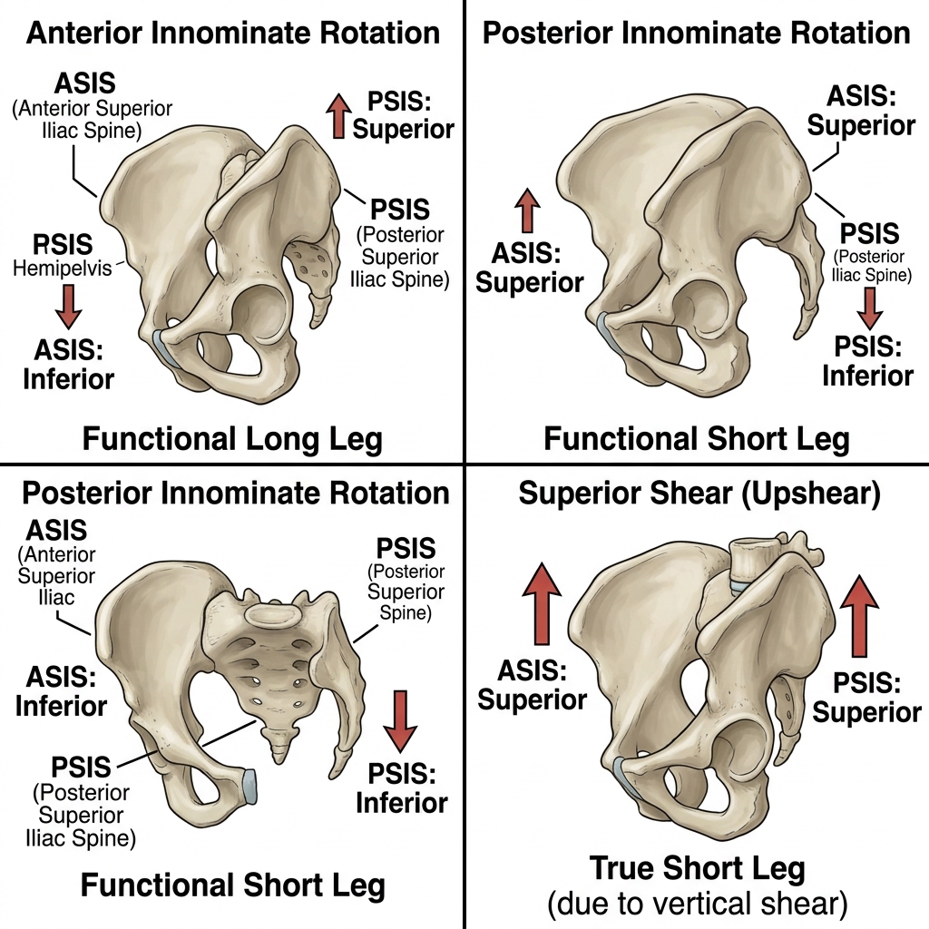
  3. 3. ASIS & PSIS Palpation: If Standing Flexion is positive, assess anterior vs. posterior rotation, superior vs. inferior shear, and inflare vs. outflare. Anterior Rotation: ASIS inferior, PSIS superior on positive side; Posterior Rotation: ASIS superior, PSIS inferior on positive side.
  4. 4. Sacral Sulcus & Inferior Lateral Angle (ILA) Palpation: Palpate bilateral sacral sulci (deep vs. shallow) and ILAs (posterior/inferior vs. anterior/superior).
  5. 5. Spring Test & Sphinx Test (Lumbosacral Spring): Assess sacral base motion in extension. Poor spring / worsening asymmetry in prone extension indicates backward sacral torsion or bilateral sacral extension (sacral base stuck posterior). Good spring / improved symmetry in prone extension indicates forward sacral torsion or bilateral sacral flexion.
  6. 6. Synthesize Diagnosis: Combine seated flexion, sulcus depth, ILA position, and spring test to name sacral torsions (e.g., Right-on-Left backward torsion vs. Left-on-Left forward torsion).

Fryette Laws & Spinal Motion Restriction Diagnosis

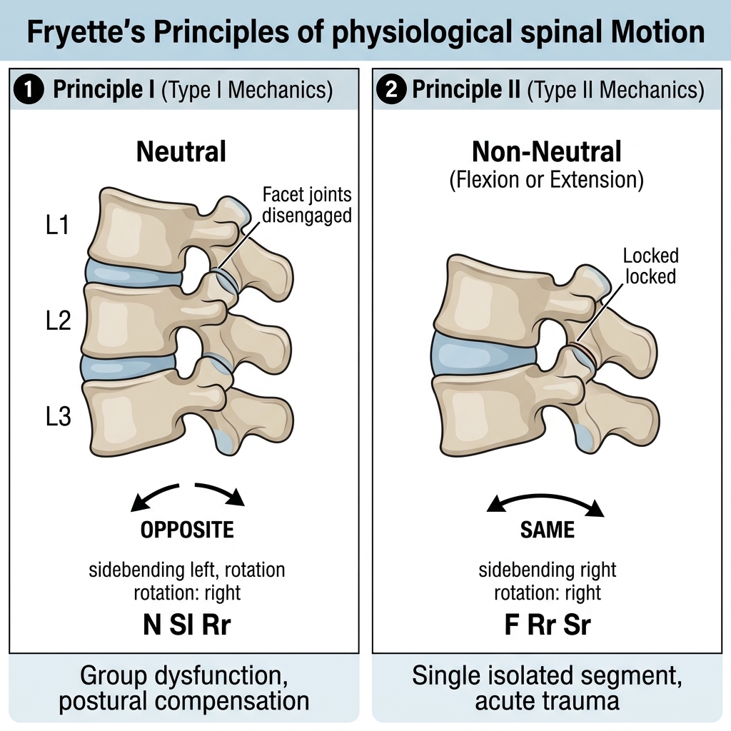
  1. 1. Neutral Positioning (Fryette Law I): Thoracic and lumbar spine in neutral (no flexion/extension). Rotation and sidebending occur to OPPOSITE sides (e.g., T4 N Sl Rr). Involves groups of 3 or more vertebrae; compensatory.
  2. 2. Non-Neutral Positioning (Fryette Law II): Thoracic and lumbar spine in marked Flexion or Extension. Rotation and sidebending occur to the SAME side (e.g., L3 F Rr Sr). Involves a single isolated segment; traumatic.
  3. 3. Principle III (Nelson's Law): Motion of a spinal segment in any one plane (flexion/extension, rotation, sidebending) reduces and restricts available motion in all other planes.
  4. 4. Cervical Exceptions: C0–C1 (Occipitoatlantal): Sidebending and rotation occur to OPPOSITE sides (like Type I). C1–C2 (Atlantoaxial): Pure ROTATION (50% of total cervical rotation; minimal sidebending/flexion). C2–C7: Sidebending and rotation occur to the SAME side in all positions (like Type II).

Stepwise OMT Contraindications & Red Flag Protocol

  1. 1. Patient Consent & Comfort: Confirm verbal consent, ensure proper positioning, and establish mutual trust before initiating any palpatory or manipulative treatment.
  2. 2. Screen for Red Flags: Evaluate for unreduced fractures, severe acute disc herniations with progressive neurologic deficit, acute spinal cord compression/cauda equina, fever with spinal tenderness (epidural abscess/osteomyelitis), or visceral emergencies (peritonitis, aneurysm rupture).
  3. 3. Modality Selection: Direct vs. Indirect. Direct techniques (Muscle Energy, HVLA) carry the restriction barrier towards ease and engage the barrier; Indirect techniques (Counterstrain, Facilitated Positional Release) move away from the barrier into the position of ease.
  4. 4. Post-Treatment Re-Evaluation: Always re-test the primary diagnostic landmarks (TART criteria, range of motion, tenderpoints) immediately following treatment to verify therapeutic resolution.
Section 03 • Clinical Chapters & Active Recall

High-Yield Clerkship Review by System

Read structured textbook-style disease summaries or test yourself with active-recall flashcards across OPP / OMM clinical systems.

Showing 7 of 7 continuous textbook chapters
Chapter 1 • Somatic Dysfunction & Cervical Spine
16 min TOC

Somatic Dysfunction Foundations & Cervical Mechanics

TART Criteria, Fryette Exceptions, OA/AA/C2-C7 Biomechanics & Whiplash

Clinical Overview & Board Focus

Somatic dysfunction is impaired or altered function of related components of the somatic (body framework) system: skeletal, arthrodial, and myofascial structures, and related vascular, lymphatic, and neural elements. The four diagnostic criteria are summarized by TART: Tissue texture abnormality, Asymmetry, Restriction of motion, and Tenderness. Cervical spine biomechanics are divided into three distinct functional zones: the occipitoatlantal (OA) joint, the atlantoaxial (AA) joint, and the typical cervical segments (C2–C7).

1.1 The TART Framework & Anatomical vs. Pathological Barriers

A diagnosis of somatic dysfunction requires palpatory identification of at least two of the four TART criteria: 1. Tissue texture abnormality: Palpable changes in skin, fascia, and musculature. In acute dysfunctions, tissues feel warm, boggy, erythematous, and hypertonic, with sharp, well-localized pain. In chronic dysfunctions, tissues feel cool, pale, dry, ropy, stringy, and fibrotic, with dull ache; 2. Asymmetry: Unequal anatomical landmarks compared bilaterally (e.g., prominent transverse processes, unlevel ASIS/PSIS); 3. Restriction of motion: Impaired active or passive mobility across one or more planes of motion; and 4. Tenderness: Pain produced during palpation of a somatic structure.

Joint motion is bounded by specific physiological and mechanical barriers. The Physiological Barrier is the limit of active, voluntary range of motion produced by patient muscle effort. The Anatomical Barrier is the ultimate structural limit of passive motion imposed by bones, joint capsules, and ligaments; exceeding this barrier causes anatomical disruption (fracture, dislocation, torn ligament). In somatic dysfunction, a Pathological (Restrictive) Barrier develops within the normal physiological range of motion, prematurely arresting motion and shifting the neutral point toward the direction of ease. Osteopathic manipulative treatment (OMT) engages the restrictive barrier (direct) or moves away from it (indirect) to eliminate the pathological restriction and restore normal range.

Naming Conventions in Somatic Dysfunction
COMLEX & OPP Board Integration
Somatic dysfunctions are ALWAYS named for the direction of ease (the direction the structure moves most freely), which is the opposite of the restrictive barrier. For example, if T5 rotates easily to the right but is restricted when rotating to the left, the diagnosis is T5 Rotated Right (freedom = right, restriction = left).

1.2 Biomechanics of the Cervical Spine: OA, AA & Typical C2–C7

The cervical spine consists of three distinct biomechanical units: 1. Occipitoatlantal (OA) Joint: The condyles of the occiput articulate with the superior articular facets of C1 (atlas). The primary motion is Flexion and Extension ('nodding' motion), accounting for 50% of total cervical flexion/extension. Crucially, Sidebending and Rotation occur to OPPOSITE sides, behaving like Fryette Type I mechanics in both flexion and extension. 2. Atlantoaxial (AA) Joint: The inferior articular facets of C1 articulate with C2 (axis) around the odontoid process (dens). The primary motion is pure Rotation, providing 50% of total cervical rotation (approximately 45° in each direction). Sidebending and flexion/extension are negligible. To isolate and examine the AA joint, the examiner fully flexes the cervical spine to 45 degrees (locking facet joints from C2 down) and assesses passive rotation right and left.

3. Typical Cervical Segments (C2 through C7): Guided by superior articular facets that face backward, upward, and medially (BUM), sidebending and rotation are coupled to the SAME side in all positions. Lateral translation (transverse gliding) is used to assess sidebending: translating a vertebra to the right induces left sidebending; translating to the left induces right sidebending.

Whiplash (Acceleration-Deceleration Injury): Common following rear-end collisions. Rapid hyperextension causes severe strain of the anterior cervical muscles (longus colli, longus capitis, sternocleidomastoid, scalenes), followed by rebound hyperflexion causing posterior ligamentous strain. Treat acutely with gentle indirect modalities (counterstrain, myofascial release) to prevent persistent post-traumatic cervical lordosis loss.
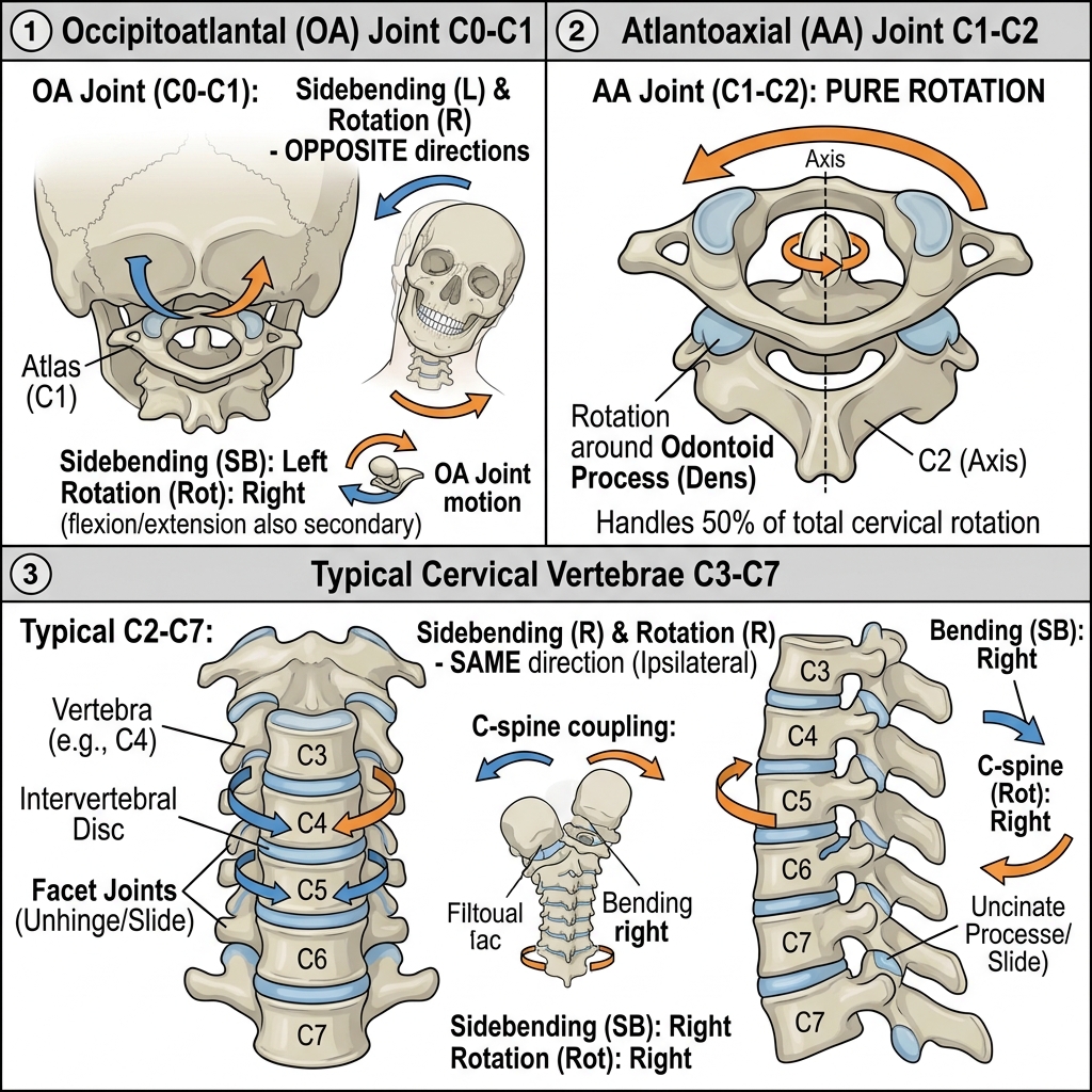
Cervical Spine Biomechanics and Facet Orientations
Diagram illustrating the BUM facet orientation (Backward, Upward, Medial) of the typical cervical spine (C2–C7) dictating coupled rotation and sidebending to the same side.

Chapter 1 Quick-Check Self-Assessment

Test your clinical reasoning before moving to the next chapter
1 Vignettes
Board Vignette #1 Single Best Answer
A patient presents with limited cervical rotation. Examination reveals that with the neck flexed to 45 degrees, rotation to the right is 20 degrees and rotation to the left is 45 degrees. What is the diagnosis?
Estimated study time: 16 min
Chapter 2 • Cranial Osteopathy & PRM
17 min TOC

Osteopathy in the Cranial Field & Primary Respiratory Mechanism

Five Components of PRM, Sphenobasilar Synchondrosis (SBS) Strains & Cranial Nerve Entrapment

Clinical Overview & Board Focus

Osteopathy in the Cranial Field, developed by William Garner Sutherland, DO, is predicated on the Primary Respiratory Mechanism (PRM). Board questions focus on the five components of the PRM, the cranial flexion/extension cycle, distinguishing physiological (torsion, sidebending/rotation) from non-physiological (vertical, lateral, compression) SBS strain patterns, and cranial nerve compression syndromes.

2.1 The Five Components of the Primary Respiratory Mechanism (PRM)

The Primary Respiratory Mechanism operates continuously throughout life independent of thoracic pulmonary respiration. Sutherland identified five fundamental anatomical components: 1. Inherent motility of the brain and spinal cord: The central nervous system undergoes a rhythmic, coiled pulsatile motion; 2. Fluctuation of the cerebrospinal fluid (CSF): Rhythmic hydrodynamic pulsation of CSF; 3. Mobility of the reciprocal tension membranes (RTM): The intracranial and intraspinal dura mater (falx cerebri, tentorium cerebelli, and spinal dura anchored firmly at foramen magnum, C2, C3, and S2) acts as a dynamic tension balance system; 4. Articular mobility of the cranial bones: Sutures remain microscopically mobile throughout adult life; and 5. Involuntary mobility of the sacrum between the ilia: The sacral base moves around a superior transverse axis at S2 linked to the cranium via the dural tube.

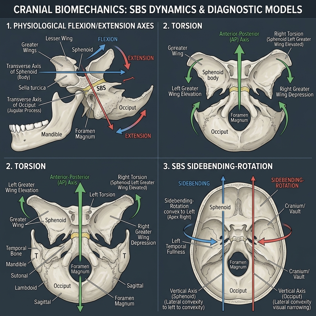
The Cranial Flexion & Extension Cycle: The Cranial Rhythmic Impulse (CRI) oscillates at 10 to 14 cycles per minute. During Cranial Flexion: The sphenobasilar synchondrosis (SBS) flexes (elevates); midline bones (sphenoid, occiput, ethmoid, vomer) flex; paired bones (temporals, parietals, maxillae) externally rotate; the transverse diameter of the skull widens while AP diameter decreases; and the sacral base moves posteriorly (cranial extension / counternutation). During Cranial Extension: The SBS drops; paired bones internally rotate; the head narrows transversely and elongates AP; and the sacral base moves anteriorly (cranial flexion / nutation).

Cranial Strain Patterns and SBS Axes
Diagrammatic representation of sphenobasilar synchondrosis (SBS) motion during cranial flexion, extension, torsions, and vertical strains.

2.2 SBS Strain Patterns & Diagnostic Vault Hold

SBS strain patterns are palpated using the Vault Hold: index finger on greater wing of sphenoid, middle finger anterior to ear on temporal zygomatic process, ring finger on temporal mastoid process, and little finger on squamous occiput. Strains are classified into physiological and non-physiological: 1. Torsion: Sphenoid and occiput rotate in opposite directions around 1 AP axis. Named for the greater wing of the sphenoid that is superior (e.g., Right Torsion: right index finger moves superiorly while right pinky moves inferiorly). 2. Sidebending / Rotation: Sphenoid and occiput rotate in opposite directions around 2 vertical axes (sidebending) and rotate in the same direction around 1 AP axis (rotation). Named for the side that feels wide and full (the low side / side of concavity).

Non-Physiological Strains: 1. Vertical Strain: Sphenoid and occiput rotate in the same direction around 2 transverse axes. Named for the direction of the sphenoid base: Superior vertical strain (sphenoid base moves up; index fingers move down) or Inferior vertical strain. 2. Lateral Strain: Sphenoid and occiput rotate in the same direction around 2 vertical axes, creating a 'parallelogram-shaped' head. Named for the direction of the sphenoid base (Right or Left). 3. Compression: The sphenoid and occiput are driven together, severely restricting the CRI; the head feels rigid and dense like a bowling ball, commonly linked to severe clinical depression or traumatic birth.

Condylar Compression & Infant Poor Suckling
COMLEX & OPP Board Integration
Birth trauma from prolonged labor or forceps delivery can compress the occipital condyles against the lateral masses of C1, impinging Cranial Nerve XII (Hypoglossal) in the hypoglossal canal. Neonates present with poor suckling, difficulty latching onto the breast, and irritability. Gentle condylar decompression releases the occipital squama from the condyles and immediately restores normal tongue motor function and latch.

Chapter 2 Quick-Check Self-Assessment

Test your clinical reasoning before moving to the next chapter
1 Vignettes
Board Vignette #1 Single Best Answer
During the vault hold examination, the physician's right index finger moves superiorly while the right fifth digit moves inferiorly. The left index finger moves inferiorly and left fifth digit moves superiorly. What is the diagnosis?
Estimated study time: 17 min
Chapter 3 • Thorax, Ribs & Lymphatics
16 min TOC

Thoracic Spine, Rib Mechanics & Lymphatic Circulatory Models

Pump-Handle vs. Bucket-Handle vs. Caliper, Inhalation/Exhalation Key Ribs & Sibson's Fascia

Clinical Overview & Board Focus

Thoracic cage mobility directly governs cardiovascular venous return, lymphatic circulation, and pulmonary ventilation. Board exams test rib motion kinematics (pump-handle, bucket-handle, caliper), the 'Key Rib' rules for group inhalation and exhalation dysfunctions, and the step-by-step sequence of terminal lymphatic drainage.

3.1 Kinematics of Respiration: Pump-Handle, Bucket-Handle & Caliper

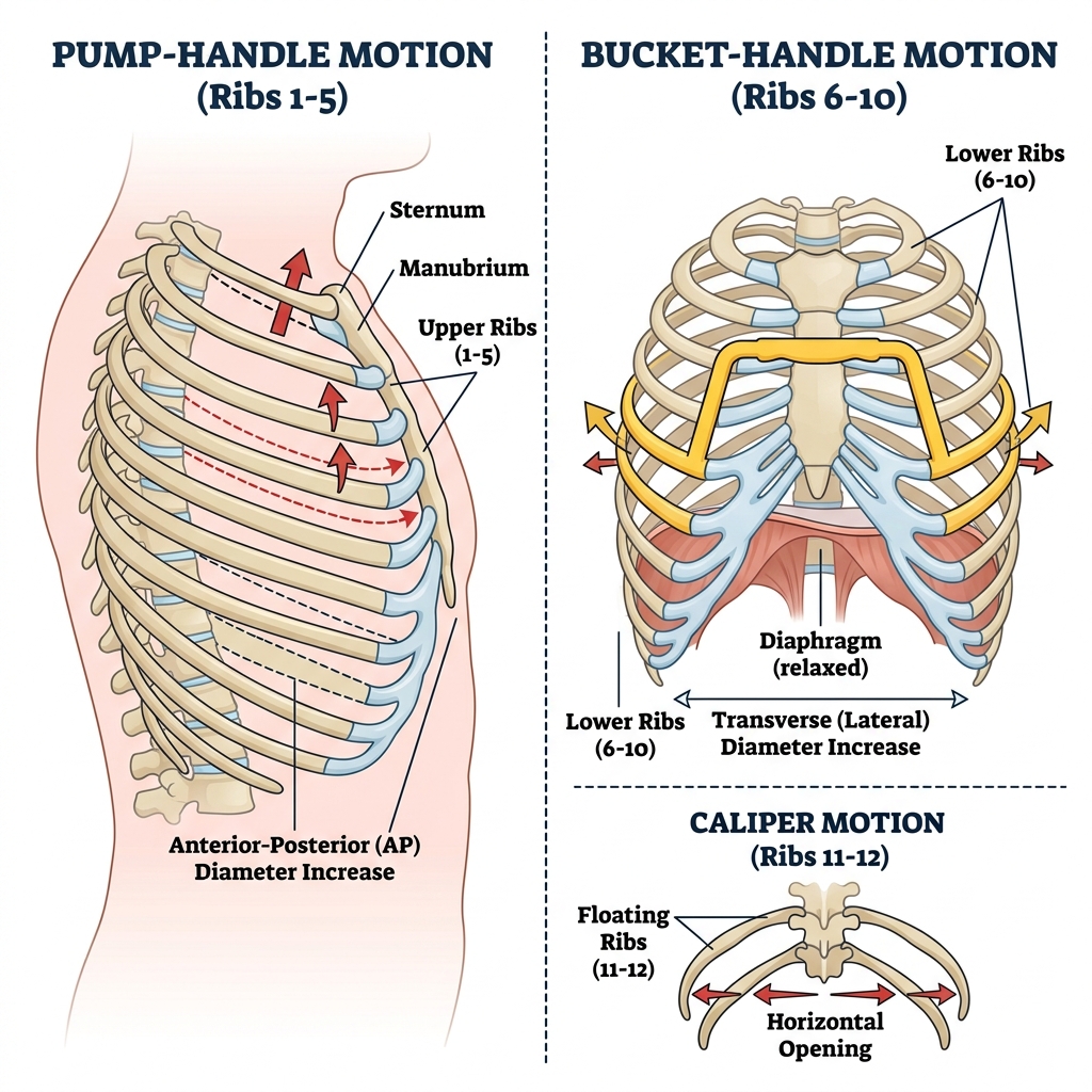
Rib motion during respiration is partitioned into three distinct biomechanical mechanisms based on anatomical level: 1. Ribs 1 through 5 (Pump-Handle Motion): Rotate primarily around a coronal/transverse axis passing through the costovertebral and costotransverse joints. During inhalation, the anterior rib ends and sternum move anteriorly and superiorly, increasing the anteroposterior (AP) diameter of the thoracic cage. 2. Ribs 6 through 10 (Bucket-Handle Motion): Rotate around an anteroposterior (sagittal) axis connecting the posterior costovertebral joint to the anterior chondrosternal junction. During inhalation, the lateral shafts of the ribs swing laterally and superiorly, increasing the transverse diameter of the thorax. 3. Ribs 11 and 12 (Caliper Motion): Floating ribs with no anterior sternal attachment or costotransverse joints. During inhalation, they move posterior, inferior, and laterally (caliper-like opening) around a vertical axis.

Pump-Handle vs. Bucket-Handle Rib Motion
Diagram illustrating pump-handle motion (ribs 1–5, increasing AP diameter) and bucket-handle motion (ribs 6–10, increasing transverse diameter).

3.2 Inhalation vs. Exhalation Somatic Dysfunctions & Key Rib Rules

Rib dysfunctions rarely occur in isolation; they typically present as a group restriction. Inhalation Somatic Dysfunction: The rib group moves freely into inhalation but is restricted in exhalation (ribs locked up). The Key Rib is the LOWEST rib in the group ('Bottom Inhalation'). The lowest rib prevents the ribs above it from descending into exhalation. Treat the bottom rib first.

Exhalation Somatic Dysfunction: The rib group moves freely into exhalation but is restricted in inhalation (ribs locked down). The Key Rib is the TOP (HIGHEST) rib in the group ('Top Exhalation'). The top rib prevents the ribs below it from elevating into inhalation. Treat the top rib first. Motor Muscles for Muscle Energy: Rib 1 = Anterior/Middle Scalenes; Rib 2 = Posterior Scalene; Ribs 3–5 = Pectoralis Minor; Ribs 6–9 = Serratus Anterior; Ribs 10–11 = Latissimus Dorsi; Rib 12 = Quadratus Lumborum.

3.3 The Lymphatic Model & Sequence of Drainage

The lymphatic system lacks a central muscular pump, relying entirely on negative intrathoracic pressure, diaphragmatic excursion, and skeletal muscle contraction. Sequential Treatment Principle: To treat lymphatic congestion, the physician must ALWAYS treat central/proximal restrictions before distal lymphatic pumps. 1. Release the Thoracic Inlet (Sibson's Fascia): Unrestricting the superior thoracic aperture allows the thoracic duct (draining 75% of the body) and right lymphatic duct to empty freely into the subclavian veins. 2. Release the Thoracoabdominal Diaphragm: Redoming the diaphragm restores negative intrathoracic pumping pressure. 3. Release the Pelvic Diaphragm. 4. Apply Extrinsic Lymphatic Pumps: Thoracic pump, pectoral traction, pedal pump, or splenic/liver pump. Pumping against a restricted thoracic inlet increases terminal venous pressures and worsens edema.

Chapter 3 Quick-Check Self-Assessment

Test your clinical reasoning before moving to the next chapter
1 Vignettes
Board Vignette #1 Single Best Answer
A 45-year-old female presents with right-sided chest wall pain following a severe bout of bronchitis. Examination reveals ribs 3, 4, and 5 on the right are restricted in inhalation. What is the key rib that must be treated first and which muscle is engaged in Muscle Energy?
Estimated study time: 16 min
Chapter 4 • Lumbar Spine & Fryette Mechanics
16 min TOC

Lumbar Spine Biomechanics, Fryette's Principles & Psoas Syndrome

Type I vs Type II Mechanics, Ferguson's Angle, Spondylolisthesis & Lumbar Radiculopathy

Clinical Overview & Board Focus

The lumbar spine bears the greatest axial load of the vertebral column. Mastery of Harrison Fryette's principles of spinal motion, the clinical presentation and multi-step progression of Psoas Syndrome, Ferguson's lumbosacral angle, and differentiating neurogenic claudication from vascular disease are essential board topics.

4.1 Fryette's Principles of Physiological Spinal Motion

In 1918, Harrison Fryette, DO, formulated the biomechanical laws of thoracic and lumbar spinal motion. Principle I: When the thoracic or lumbar spine is in Neutral (neither marked flexion nor extension), sidebending and rotation occur to OPPOSITE sides (e.g., L1–L4 N Sl Rr). Type I dysfunctions involve groups of three or more consecutive vertebrae, maintained by long superficial polyarticular muscles (erector spinae), and represent postural compensation.

Principle II: When the spine is in Non-Neutral (marked Flexion or Extension), sidebending and rotation occur to the SAME side (e.g., L3 F Rr Sr). Type II dysfunctions involve a single isolated vertebral segment, maintained by deep monoarticular muscles (rotatores, intertransversarii, multifidi), and typically result from acute traumatic strain.

Principle III (Nelson's Law): Initiating motion of a spinal segment in any one plane reduces and restricts its available motion in the other two planes.

Lumbar Spine Fryette Type I and Type II Biomechanics
Biomechanical diagram illustrating Type I (neutral, sidebending and rotation opposite) vs. Type II (non-neutral, sidebending and rotation to same side) in the lumbar spine.

4.2 Psoas Syndrome & Lumbosacral Pathology

The iliopsoas originates from T12–L5 vertebral bodies, transverse processes, and discs, inserting onto the lesser trochanter of the femur. Psoas Syndrome typically begins after prolonged hip flexion (sitting, driving) followed by sudden extension. Spasm of the psoas initiates a classic sequence: 1. A key Type II somatic dysfunction at L1 or L2 flexed, rotated, and sidebent to the side of the hypertonic psoas; 2. Pelvic shift to the OPPOSITE side; 3. A functionally short leg with foot externally rotated on the affected side; 4. A compensatory contralateral piriformis spasm (which can produce sciatic nerve irritation); and 5. A positive Thomas test on the affected side.

Spondylolysis and Spondylolisthesis: Spondylolysis is a stress fracture of the pars interarticularis (most commonly L5-S1), appearing as a radiolucent 'collar on the Scotty dog' on oblique lumbar radiographs. When bilateral pars defects allow anterior subluxation of the superior vertebral body, Spondylolisthesis occurs ('decapitated Scotty dog'). Ferguson's angle (normal 25–35°) is increased, amplifying anterior shear forces.

Chapter 4 Quick-Check Self-Assessment

Test your clinical reasoning before moving to the next chapter
1 Vignettes
Board Vignette #1 Single Best Answer
A patient presents with acute low back pain. Examination reveals L2 is restricted in extension and right rotation. When fully flexed, the right transverse process is markedly posterior compared to the left; in extension, the asymmetry worsens. What is the diagnosis?
Estimated study time: 16 min
Chapter 5 • Sacrum & Innominate Torsions
18 min TOC

Sacral Torsions, Shears & Innominate Rotations

L5/Sacrum Rules, Forward vs Backward Torsions, Spring/Sphinx Tests & Pelvic Shears

Clinical Overview & Board Focus

Sacral and pelvic somatic dysfunctions represent the highest-yield, most heavily tested topic on the COMLEX and COMAT OPP examinations. Clear differentiation between sacroiliac (sacrum on ilium) and iliosacral (ilium on sacrum) mechanics, mastery of the Fred Mitchell sacral oblique axes, and the universal L5/Sacrum relationship guarantees board mastery.

5.1 Sacral Oblique Axes & Forward vs. Backward Torsions

Sacral torsions occur around two diagonal Oblique Axes named for the superior pole from which they originate: the Right Oblique Axis runs from the right superior sacral base to the left inferior lateral angle (ILA); the Left Oblique Axis runs from the left superior sacral base to the right ILA. Torsions are named as: [Sacral Rotation] on [Oblique Axis] (e.g., Left-on-Left or Right-on-Left).

The Seated Flexion Test: Assesses sacroiliac motion. The side of the positive seated flexion test (where the PSIS moves further superiorly) is OPPOSITE the oblique axis. If seated flexion is positive on the right, the axis is the Left Oblique Axis; if positive on the left, the axis is the Right Oblique Axis.

Spring Test & Sphinx Test: Assess whether a torsion is Forward (physiological) or Backward (non-physiological). A Negative Spring Test (good spring / resilient end-feel) and a Positive Sphinx Test (asymmetry improves when patient prows up on elbows into extension) indicates a Forward Torsion (L-on-L or R-on-R). A Positive Spring Test (poor spring / hard, rigid stop) and a negative Sphinx test (asymmetry worsens in extension) indicates a Backward Torsion (R-on-L or L-on-R).

Sacral Torsion Diagnosis Algorithm
Diagnostic algorithm for sacral torsions integrating seated flexion test, sulcus depth, ILA position, and spring/Sphinx test results.

5.2 Innominate Rotations, Shears & Pubic Dysfunctions

Standing Flexion Test: Assesses iliosacral motion (innominate moving on sacrum). The side on which the PSIS travels further superiorly is the positive side (side of innominate dysfunction). 1. Anterior Innominate Rotation: ASIS is inferior and PSIS is superior on the positive side; associated with a functionally longer leg; treated with Muscle Energy engaging the ipsilateral hamstrings. 2. Posterior Innominate Rotation: ASIS is superior and PSIS is inferior on the positive side; associated with a functionally shorter leg; treated with Muscle Energy engaging the ipsilateral rectus femoris/quadriceps.

3. Superior Innominate Shear (Upshear): Both the ASIS and PSIS are superior on the positive standing flexion side, with a superior pubic tubercle. Caused by sudden vertical trauma (stepping into a hole or dashboard impact). 4. Pubic Shear: Superior or inferior displacement of one pubic ramus relative to the other at the pubic symphysis, commonly producing groin and lower abdominal pain in postpartum patients.

Innominate Rotations and Pelvic Shears
Pelvic landmarks demonstrating Anterior Rotation (ASIS inferior, PSIS superior) and Posterior Rotation (ASIS superior, PSIS inferior).

Chapter 5 Quick-Check Self-Assessment

Test your clinical reasoning before moving to the next chapter
1 Vignettes
Board Vignette #1 Single Best Answer
A 32-year-old runner has a positive seated flexion test on the left. The right sacral sulcus is deep and the left ILA is posterior/inferior. The spring test demonstrates good spring/resilience at the lumbosacral junction. What is the diagnosis?
Estimated study time: 18 min
Chapter 6 • Viscerosomatics & Chapman Points
17 min TOC

Viscerosomatic Reflexes, Autonomic Mapping & Chapman Points

Sympathetic/Parasympathetic Ganglia, Clinical Chapman Points & Autonomic Normalization

Clinical Overview & Board Focus

Viscerosomatic reflexes occur when visceral disease produces somatic sensory, motor, and trophic changes at spinal segments sharing the same sympathetic or parasympathetic innervation. Mastery of collateral sympathetic ganglia levels (Celiac, SMG, IMG), the Vagus vs. Pelvic Splanchnic transition, and the exact anatomical locations of Chapman reflex points guarantees top scores on COMLEX exams.

6.1 Autonomic Levels & The Three Collateral Sympathetic Ganglia

Sympathetic preganglionic neurons originate strictly in the intermediolateral cell column (IML) from T1 to L2. 1. Celiac Ganglion (T5–T9): Innervates foregut structures: distal esophagus, stomach, liver, gallbladder, spleen, portions of pancreas, and 1st/2nd duodenum. 2. Superior Mesenteric Ganglion (T10–T11): Innervates midgut structures: 3rd/4th duodenum, jejunum, ileum, ascending colon, proximal 2/3 of transverse colon, appendix, kidneys, and gonads (ovaries/testes). 3. Inferior Mesenteric Ganglion (T12–L2): Innervates hindgut and pelvic structures: distal 1/3 of transverse colon, descending colon, sigmoid colon, rectum, lower ureters, bladder, and reproductive organs (uterus, prostate).

Parasympathetic Division: 1. Vagus Nerve (CN X): Emerges through the jugular foramen to supply all viscera from the occiput down to the splenic flexure (distal 2/3 transverse colon), as well as the kidneys and upper ureters. 2. Pelvic Splanchnic Nerves (S2–S4): Arise from sacral spinal cord to innervate the distal 1/3 of the transverse colon, descending colon, sigmoid, rectum, bladder, and reproductive organs.

Viscerosomatic Reflex Pathways and Chapman Points
Anatomical map correlating spinal autonomic segments (T1–L2) with visceral organ reflex manifestations and anterior/posterior Chapman points.

6.2 High-Yield Chapman Reflex Points

Chapman points are small, discrete, pea-sized fibroelastic nodules located in the deep fascia, representing localized visceral-sympathetic-lymphatic reflexes. High-Yield Anterior Points: 1. Heart: 2nd intercostal space adjacent to the sternum; 2. Lungs: 3rd and 4th intercostal spaces adjacent to the sternum; 3. Stomach: Left 6th intercostal space between midclavicular line and sternum; 4. Liver & Gallbladder: Right 6th intercostal space; 5. Pancreas: Right 7th intercostal space; 6. Appendix: Tip of the right 12th rib anteriorly; 7. Kidneys: 1 inch superior and 1 inch lateral to umbilicus; 8. Adrenals: 2 inches superior and 1 inch lateral to umbilicus; 9. Colon: Lateral aspect of the Iliotibial (IT) band from greater trochanter to knee.

Chapter 6 Quick-Check Self-Assessment

Test your clinical reasoning before moving to the next chapter
1 Vignettes
Board Vignette #1 Single Best Answer
A 22-year-old male presents with acute appendicitis. At which spinal levels would you expect to find an acute viscerosomatic tissue texture abnormality, and where is the anterior Chapman point?
Estimated study time: 17 min
Chapter 7 • OMT Modalities & Orthopedic Tests
17 min TOC

Treatment Modalities, Counterstrain & Orthopedic Special Tests

Direct vs Indirect, Muscle Energy, Counterstrain Tenderpoints, HVLA Rules & Extremity Tests

Clinical Overview & Board Focus

Selecting the optimal treatment modality requires understanding patient indications, acute versus chronic pathology, and absolute contraindications. Direct modalities (HVLA, Muscle Energy) engage the barrier, whereas indirect modalities (Counterstrain, FPR) move away from the barrier into the position of ease. High-frequency orthopedic special tests evaluate peripheral joint instability.

7.1 Modality Comparison: Direct vs. Indirect & Neurophysiology

Muscle Energy Technique (MET): An active, direct technique invented by Fred Mitchell, Sr., DO. The physician engages the restrictive barrier across all three planes, and the patient performs an isometric contraction against unyielding counterforce for 3 to 5 seconds. Following complete relaxation, the physician takes up the slack to the new barrier. Mediated by Golgi Tendon Organs (post-isometric relaxation).

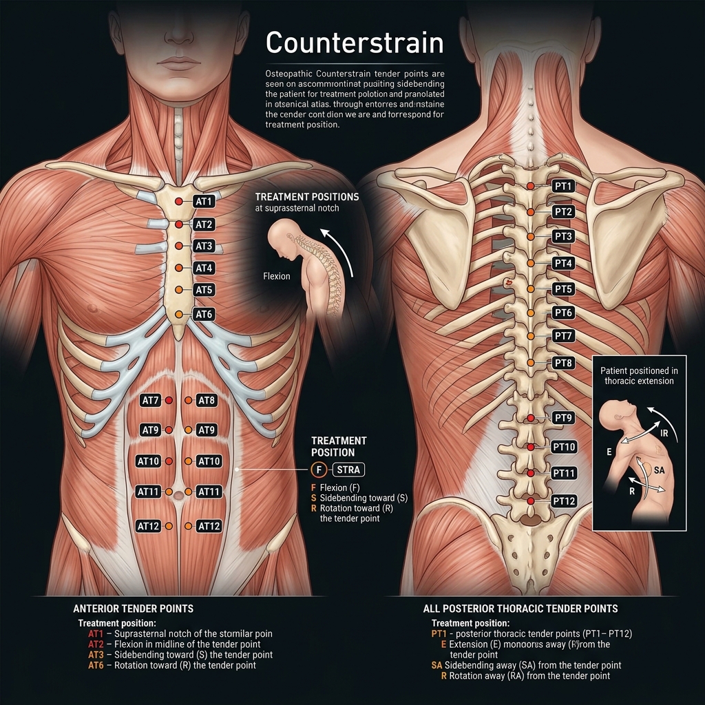
Counterstrain (Jones Technique): A passive, indirect technique developed by Lawrence Jones, DO. The physician identifies an exquisitely tender point (10/10 pain), positions the patient away from the barrier into the position of ease until tenderness decreases by at least 70% (down to <= 3/10), maintains the position for 90 seconds (120 seconds for ribs), and slowly, passively returns the patient to neutral. Mediated by muscle spindle silencing.

High-Velocity Low-Amplitude (HVLA): A passive, direct technique utilizing a rapid, short-distance therapeutic thrust through the restrictive barrier. Absolute contraindications: Fractures, acute dislocations, RA/Down syndrome cervical instability, bone tumors, osteomyelitis, and patient refusal.

Counterstrain Thoracic Tenderpoints
Anatomical map of anterior thoracic (AT1–AT12) and posterior thoracic tenderpoint locations with their corresponding flexion and extension treatment setups.

7.2 Orthopedic Special Tests for Board Exams

1. Shoulder Tests: Neer & Hawkins: Subacromial impingement; Empty Can (Jobe): Supraspinatus tear; Speed & Yergason: Biceps tendinitis; Apprehension Test: Anterior glenohumeral instability.

2. Knee Tests: Lachman (most sensitive at 30°) & Anterior Drawer: ACL tear; Posterior Drawer & Sag Sign: PCL tear; McMurray & Thessaly: Meniscal tears; Valgus / Varus Stress: MCL / LCL integrity.

3. Hip & Pelvis Tests: FABER (Patrick): Pain anteriorly = hip joint arthritis; pain posteriorly = sacroiliac joint; Thomas Test: Psoas contracture; Trendelenburg Test: Gluteus medius weakness (superior gluteal nerve).

Orthopedic Special Tests for Hip and Pelvis
Illustration of FABER (Patrick), Thomas, and Trendelenburg physical exam tests for diagnosing sacroiliac and lumbopelvic pathology.

Chapter 7 Quick-Check Self-Assessment

Test your clinical reasoning before moving to the next chapter
1 Vignettes
Board Vignette #1 Single Best Answer
A physician is treating an acute tenderpoint on the right piriformis muscle using Counterstrain. Which of the following describes the ideal therapeutic position?
Estimated study time: 17 min