Skip to content
NBOME Obstetrics & Gynecology COMAT & Level 2-CE NBME Obstetrics & Gynecology Shelf Exam & Step 2 CK

Obstetrics & Gynecology COMAT Command Center Obstetrics & Gynecology Shelf Exam Command Center

Comprehensive women's health, labor & delivery, and gynecologic oncology guide for NBOME Obstetrics & Gynecology COMAT and COMLEX Level 2-CE. Ingests 1,870+ cards covering prenatal screening, hypertensive disorders of pregnancy, abnormal uterine bleeding, and obstetric OMM.

254 Cards Extracted 2 High-Yield Protocols 2 Osteopathic Rules 4 High-Yield Pearls
Section 01 • Osteopathic Principles

"Don't Miss" COMAT Osteopathic Pearls

High-frequency somatic dysfunctions, viscerosomatics, autonomic reflexes, and treatment rules

Tested heavily on NBOME Shelf

Obstetric Biomechanics Across Trimesters

As the gravid uterus expands, the center of gravity shifts anteriorly, resulting in progressive lumbar lordosis, anterior pelvic tilt, and compensatory thoracic kyphosis. Elevated relaxin softens sacroiliac ligaments. Safe techniques: Gentle muscle energy, myofascial release, and indirect counterstrain. Avoid high-velocity low-amplitude (HVLA) thrusts to the pelvis during the 3rd trimester.

Uterine & Ovarian Autonomics

Ovaries: T10–T11 sympathetics; Vagus parasympathetics. Uterus: T12–L2 sympathetics; Pelvic splanchnics (S2–S4) parasympathetics. S2–S4 sacral rocking promotes cervical dilation and eases labor discomfort. CV4 technique stimulates rhythmic CSF flow and encourages coordinated uterine contractions in post-term pregnancy.

Section 01 • Clinical Foundations

"Don't Miss" Obstetrics & Gynecology Clinical Pearls & Shelf Traps

High-frequency diagnostic pitfalls, gold-standard criteria, and next-best-step clinical rules

Tested heavily on NBME Shelf & Step 2 CK

Third-Trimester Bleeding: Placenta Previa vs. Placental Abruption

Placenta Previa: Painless bright red vaginal bleeding in 3rd trimester; NEVER perform digital vaginal exam (risk of catastrophic exsanguinating hemorrhage)—first-line evaluation is transabdominal ultrasound followed by transvaginal ultrasound. Placental Abruption (Abruptio Placentae): Painful dark red vaginal bleeding, hypertonic 'woody' tender uterus, maternal hypertension/cocaine risk factor; high risk of Disseminated Intravascular Coagulation (DIC) and fetal demise; deliver emergently if unstable or in fetal distress.

Preeclampsia Diagnostic Criteria & Acute Hypertensive Crisis

Preeclampsia requires new-onset BP >= 140/90 after 20 weeks gestation PLUS proteinuria (>= 300 mg/24h or U-Prot/Cr >= 0.3) OR severe features (BP >= 160/110, platelets < 100k, Cr > 1.1, transaminases 2x normal, pulmonary edema, new visual symptoms). Immediate seizure prophylaxis: IV Magnesium Sulfate (4–6 g IV bolus, 1–2 g/hr infusion; antidote is Calcium Gluconate 1 g IV). Acute BP control: IV Labetalol (avoid in bradycardia/asthma) or IV Hydralazine, or oral Nifedipine.

Postpartum Hemorrhage: The 4 T's & Uterotonic Escalation

Tone (Uterine Atony, 80%): Soft boggy uterus. Step 1: Bimanual uterine massage and IV Oxytocin infusion. Step 2: Methylergonovine (Methergine—strictly contraindicated in hypertension/preeclampsia due to vasoconstriction). Step 3: Carboprost tromethamine (Hemabate, PGF2a—strictly contraindicated in asthma due to bronchospasm). Step 4: Misoprostol (PGE1). Step 5: Intrauterine balloon tamponade (Bakri balloon) or emergent uterine artery embolization / surgical ligation.

Abnormal Uterine Bleeding (AUB) PALM-COEIN Classification

Structural causes (PALM): Polyp, Adenomyosis (uniformly enlarged tender boggy uterus), Leiomyoma (fibroids, irregularly enlarged non-tender firm uterus), Malignancy/hyperplasia. Non-structural causes (COEIN): Coagulopathy (von Willebrand), Ovulatory dysfunction (PCOS, anovulatory bleeding), Endometrial, Iatrogenic (IUD, anticoagulants), Not otherwise classified. Endometrial Biopsy indications: Age >= 45 with AUB, or Age < 45 with persistent bleeding and unopposed estrogen exposure (obesity, chronic anovulation/PCOS, tamoxifen, Lynch syndrome).

Section 02 • Clinical Algorithms

Core Clinical Protocols & Diagnostic Trees

Step-by-step first-line management pathways, diagnostic thresholds, and pharmacological escalation

2 Diagnostic Algorithms

Hypertensive Disorders of Pregnancy Diagnostic Algorithm

  1. 1. Gestational Hypertension: New-onset BP >= 140/90 mmHg after 20 weeks gestation without proteinuria or signs of end-organ damage.
  2. 2. Preeclampsia: BP >= 140/90 after 20 weeks gestation PLUS proteinuria (>= 300 mg/24h or urine protein/Cr ratio >= 0.3) OR severe features.
  3. 3. Severe Features: BP >= 160/110 mmHg on two occasions >= 4h apart, platelets < 100,000/uL, serum Cr > 1.1 mg/dL, AST/ALT > 2x normal, pulmonary edema, or new-onset persistent headache/visual scotoma.
  4. 4. Management: Seizure prophylaxis with IV Magnesium Sulfate (4-6 g loading, 1-2 g/h maintenance; monitor patellar reflexes, urine output, RR; calcium gluconate is antidote). Antihypertensives for severe BP: IV Labetalol, IV Hydralazine, or oral Nifedipine.
  5. 5. Delivery: At 37 weeks for preeclampsia without severe features; at 34 weeks (or immediately if unstable) for preeclampsia with severe features.

Third-Trimester Vaginal Bleeding Diagnostic Tree

  1. 1. INITIAL MANDATORY STEP: Transabdominal ultrasound BEFORE any digital vaginal examination to exclude placenta previa.
  2. 2. Placenta Previa: Painless bright red vaginal bleeding; placenta covers internal cervical os. Avoid digital exam; perform Cesarean delivery at 36-37 weeks (or emergently if severe bleeding).
  3. 3. Placental Abruption: Painful dark red vaginal bleeding with persistent uterine hypertonicity/pain, fetal distress. Risk factors: Maternal hypertension, preeclampsia, cocaine use, trauma, smoking. Emergent delivery if maternal or fetal compromise.
  4. 4. Uterine Rupture: Sudden severe tearing abdominal pain, loss of fetal station, cessation of uterine contractions, maternal hemodynamic collapse. Associated with prior Cesarean hysterotomy scar. Immediate laparotomy and delivery.
Section 03 • Clinical Chapters & Active Recall

High-Yield Clerkship Review by System

Read structured textbook-style disease summaries or test yourself with active-recall flashcards across Obstetrics & Gynecology clinical systems.

Showing 9 of 9 continuous textbook chapters
Chapter 1 • Prenatal Care, Genetics & Screening
13 min TOC

Prenatal Care, Genetic Aneuploidy Screening & Teratology

First and Second Trimester Routine Testing, Cell-Free DNA, Carrier Screening, and Medication Safety in Pregnancy

Clinical Overview & Board Focus

Routine antepartum care follows a rigorous gestational timeline designed to detect maternal asymptomatic infection, isoimmunization risk, gestational diabetes, and fetal chromosomal aneuploidies. Board exams emphasize the exact gestational timing of screening tests, differentiation between screening and diagnostic genetic modalities, and drug contraindications.

1.1 Routine Antepartum Screening Protocol

The initial prenatal visit (typically 8–10 weeks) establishes baseline maternal health and gestational dating. Essential baseline labs include: complete blood count (screening for anemia), blood type and Rh antibody screen, rubella immunity IgG, varicella immunity, urine culture (screening for asymptomatic bacteriuria), syphilis serology (RPR/VDRL), HIV 4th generation antigen/antibody assay, hepatitis B surface antigen (HBsAg), and cervical Pap smear if due. In high-risk patients, baseline hemoglobin A1c and targeted urine NAAT for Chlamydia trachomatis and Neisseria gonorrhoeae are performed.

Gestational age is most accurately established by crown-rump length (CRL) on first-trimester ultrasound (accurate to within ± 5–7 days). If menstrual dating and ultrasound dating disagree by > 7 days in the first trimester, pregnancy due date (EDD) must be adjusted according to the ultrasound. Asymptomatic bacteriuria (defined as ≥ 10^5 CFU/mL of a single organism, most commonly Escherichia coli) must ALWAYS be treated in pregnant women due to progesterone-mediated ureteral dilation and smooth muscle relaxation that dramatically increases the risk of acute pyelonephritis, preterm labor, and low birth weight. Safe first-line regimens include Nitrofurantoin (avoided in 1st trimester and near term due to hemolytic anemia), Amoxicillin-Clavulanate, or Cephalexin × 5–7 days, followed by a mandatory repeat 'test of cure' urine culture.

Subsequent visits follow a strict chronological schedule: at 15–20 weeks, maternal serum quad screen (MSAFP, beta-hCG, unconjugated estriol, inhibin A) or targeted anatomic survey ultrasound (18–22 weeks) evaluates structural anatomy and neural tube defects (elevated MSAFP). At 24–28 weeks: 1-hour 50g oral glucose challenge test (threshold ≥ 130–140 mg/dL triggers 3-hour 100g diagnostic GTT), repeat CBC for physiologic dilutional anemia, and repeat Rh antibody screen. Unsensitized Rh-negative mothers receive 300 mcg of anti-D immune globulin (RhoGAM) at 28 weeks, and again within 72 hours of delivery if the infant is Rh-positive. At 36 0/7 to 37 6/7 weeks: rectovaginal culture for Group B Streptococcus (GBS) is universally obtained.

Serum Aneuploidy Screening Patterns
Clinical Matrix
Aneuploidy / ConditionMSAFPbeta-hCGUnconjugated Estriol (uE3)Inhibin AConfirmatory Modality
Down Syndrome (Trisomy 21)Decreased (↓)Elevated (↑↑)Decreased (↓)Elevated (↑↑)CVS (10–13 wk) or Amniocentesis (15+ wk)
Edwards Syndrome (Trisomy 18)Decreased (↓)Decreased (↓↓)Decreased (↓)Decreased or NormalAmniocentesis (karyotype / microarray)
Patau Syndrome (Trisomy 13)Decreased (↓)Decreased (↓)NormalNormalAmniocentesis (karyotype / microarray)
Neural Tube Defect / GastroschisisMarkedly Elevated (↑↑)NormalNormalNormalTargeted level II ultrasound + amniotic AFP/AChE
Board Trap — Cell-Free Fetal DNA (cfDNA) Limitations
High-Yield Board Trap & Alert
Cell-free DNA (cfDNA) from maternal plasma can be performed anytime ≥ 10 weeks gestation and boasts exceptional sensitivity and specificity (> 99%) for Trisomies 21, 18, and 13. However, cfDNA is strictly a SCREENING test, NOT a diagnostic test. An abnormal or high-risk cfDNA result must NEVER be used alone to justify pregnancy termination. The patient must be counseled and offered invasive diagnostic testing via Chorionic Villus Sampling (CVS, 10–13 weeks) or Amniocentesis (≥ 15 weeks) to obtain definitive fetal karyotyping.

Chapter 1 Quick-Check Self-Assessment

Test your clinical reasoning before moving to the next chapter
3 Vignettes
Board Vignette #1 Single Best Answer
A 26-year-old G1P0 at 10 weeks gestation has routine prenatal screening. Urine culture yields 10^5 CFU/mL of Escherichia coli. She is completely asymptomatic without dysuria, fever, or frequency. What is the most appropriate management?
Board Vignette #2 Single Best Answer
A 26-year-old G1P0 woman at 16 weeks gestation undergoes maternal serum quadruple screening. The results show a significantly elevated maternal serum alpha-fetoprotein (MSAFP) of 3.8 MoM (normal < 2.0). The other three serum analytes are normal. Which of the following is the most likely etiology of this isolated elevation?
Board Vignette #3 Single Best Answer
A 36-year-old primigravida at 11 weeks gestation undergoes routine first-trimester screening. Cell-free DNA (cfDNA) demonstrates high risk for Trisomy 21 (Down syndrome). What is the most appropriate next step to confirm the diagnosis definitively?
Estimated study time: 13 min
Chapter 2 • Hypertensive Disorders & Maternal Medical Complications
14 min TOC

Hypertensive Disorders of Pregnancy & Maternal Medical Illness

Gestational Hypertension, Preeclampsia with Severe Features, Eclampsia, HELLP Syndrome, and Gestational Diabetes

Clinical Overview & Board Focus

Hypertensive disorders of pregnancy remain leading causes of maternal morbidity, placental abruption, and iatrogenic preterm delivery. Mastery requires clear differentiation based on gestational age (before vs. after 20 weeks), detection of end-organ severe features, seizure prophylaxis with magnesium sulfate, and blood pressure control.

2.1 The Preeclampsia Spectrum & Eclampsia Emergency Protocols

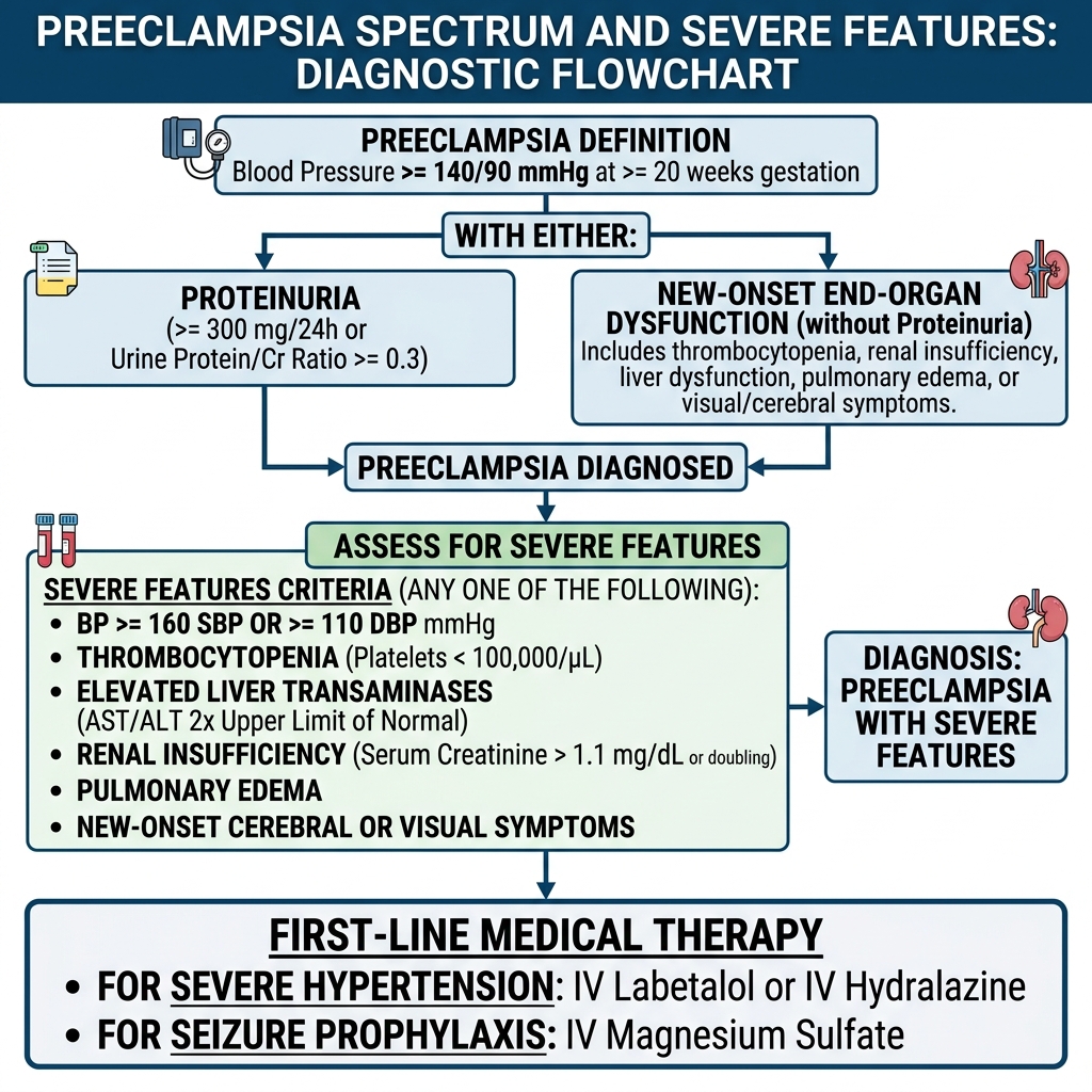
Hypertension in pregnancy is categorized chronologically and syndromically. Chronic Hypertension is defined as systolic BP ≥ 140 mmHg or diastolic BP ≥ 90 mmHg documented prior to pregnancy or before 20 weeks of gestation. Gestational Hypertension is new-onset elevated blood pressure (systolic ≥ 140 or diastolic ≥ 90 mmHg on ≥ 2 occasions at least 4 hours apart) occurring at ≥ 20 weeks gestation in the ABSENCE of proteinuria or systemic end-organ damage. Preeclampsia is diagnosed when new-onset hypertension at ≥ 20 weeks is accompanied by either Proteinuria (≥ 300 mg per 24-hour urine collection, or protein/creatinine ratio ≥ 0.3, or urine dipstick 2+) OR any 'Severe Features' in the absence of proteinuria.

Severe Features of Preeclampsia comprise: (1) Severe hypertension: systolic BP ≥ 160 mmHg or diastolic BP ≥ 110 mmHg on 2 occasions ≥ 4 hours apart while on bed rest; (2) Thrombocytopenia: platelet count < 100,000/µL; (3) Impaired liver function: transaminases (AST/ALT) ≥ 2× upper limit of normal or severe persistent right upper quadrant/epigastric pain; (4) Renal insufficiency: serum creatinine > 1.1 mg/dL or doubling of baseline; (5) Pulmonary edema; and (6) New-onset persistent cerebral or visual disturbances (scotomata, photopsia, severe refractory headache).

Management of preeclampsia without severe features is expectant until 37 0/7 weeks, at which time delivery is indicated. In preeclampsia with severe features, delivery is indicated at ≥ 34 0/7 weeks (or sooner if maternal or fetal status decompensates). Intrapartum seizure prophylaxis is administered universally using IV Magnesium Sulfate (4–6 g loading dose over 20 minutes, followed by 1–2 g/hr continuous infusion maintained through 24 hours postpartum). Acute severe hypertension (BP ≥ 160/110 mmHg sustained for 15 minutes) requires emergent IV antihypertensive therapy within 30–60 minutes: IV Labetalol (avoided in asthma or bradycardia), IV Hydralazine (risk of reflex tachycardia), or oral immediate-release Nifedipine (calcium channel blocker).

Preeclampsia Diagnostic Criteria and Management Algorithm
Diagnostic criteria and clinical management flowchart for the Preeclampsia Spectrum: Differentiating gestational hypertension from preeclampsia and identifying Severe Features (≥160/110 mmHg, platelets <100k, AST/ALT ≥2×, Cr >1.1 mg/dL, pulmonary edema, persistent cerebral/visual symptoms). Severe features mandate IV Magnesium Sulfate seizure prophylaxis and immediate blood pressure control with IV Labetalol, Hydralazine, or oral Nifedipine.
HELLP Syndrome & Magnesium Toxicity Parameters
Clinical Matrix
Clinical ParameterPathophysiology / Clinical FindingImmediate Clinical Management
HELLP SyndromeHemolysis (schistocytes, LDH > 600, indirect bili > 1.2), Elevated Liver enzymes (AST/ALT ≥ 2×), Low Platelets (< 100k)Stabilize mother, IV Magnesium sulfate, immediate delivery (regardless of gestational age if > 34 wk or unstable)
Mild Mg ToxicitySerum Mg 4.8–8.4 mg/dL: Therapeutic level; therapeutic warmth and flushingContinue infusion, monitor DTRs and hourly urine output
Moderate Mg ToxicitySerum Mg 7–10 mg/dL: Loss of deep tendon reflexes (patellar reflex disappears first)Stop Magnesium infusion immediately; obtain serum level
Severe Mg ToxicitySerum Mg 10–12 mg/dL: Respiratory depression (RR < 12/min); > 15 mg/dL: Asystole / arrestStop Mg immediately; administer IV Calcium Gluconate (10 mL of 10% solution over 2–3 min)
COMLEX Clinical Integration — Pelvic Diaphragm & Sacral Torsions in Late Pregnancy
COMLEX & OPP Board Integration
Elevated circulating relaxin and progesterone induce widespread ligamentous laxity of the pubic symphysis and sacroiliac joints. As the gravid uterus enlarges, the maternal center of gravity shifts anteriorly, inducing marked compensatory lumbar hyperlordosis and bilateral anterior sacral rotation. Paraspinal hypertonicity concentrates at the thoracolumbar junction (T10–L2, carrying uterine and renal sympathetics) and sacral base (S2–S4 parasympathetics). Gentle sacral rocking and muscle energy techniques for anterior sacral torsions relieve lumbosacral mechanical strain, enhance pelvic floor compliance, and alleviate neurovegetative congestion of the pelvic plexus.

Chapter 2 Quick-Check Self-Assessment

Test your clinical reasoning before moving to the next chapter
3 Vignettes
Board Vignette #1 Single Best Answer
A 32-year-old G2P1 at 35 weeks gestation presents with persistent severe headache and photopsia. Blood pressure is 168/112 mmHg on two readings 20 minutes apart. Urine protein/creatinine ratio is 0.8. Platelet count is 88,000/µL and AST is 142 U/L. What is the definitive management?
Board Vignette #2 Single Best Answer
A 32-year-old G1P0 woman at 34 weeks gestation with severe preeclampsia is receiving an intravenous magnesium sulfate infusion for seizure prophylaxis. Four hours into the infusion, she becomes drowsy and complains of difficulty catching her breath. Vital signs: BP 148/94 mmHg, HR 64 bpm, RR 8 breaths/min, O2 sat 90% on room air. Patellar reflexes are absent bilaterally. Which of the following is the immediate antidote?
Board Vignette #3 Single Best Answer
A 31-year-old at 34 weeks gestation presents with BP 168/112 mmHg, platelet count 72,000/uL, serum creatinine 1.3 mg/dL, and AST 184 U/L. Fetal heart tracing is Category I. In addition to IV magnesium sulfate for seizure prophylaxis, what is the most appropriate definitive management?
Estimated study time: 14 min
Chapter 3 • Labor, Delivery & Fetal Heart Monitoring
13 min TOC

Normal & Abnormal Labor, Cardiotocography & Operative Delivery

Stages of Labor, Protraction vs. Arrest Disorders, Intrapartum Fetal Heart Tracings, and Shoulder Dystocia

Clinical Overview & Board Focus

Labor management requires intimate knowledge of the active phase thresholds, cardinal movements of labor, electronic fetal monitoring categories, and emergency maneuvers for intrapartum catastrophes such as umbilical cord prolapse and shoulder dystocia.

3.1 Stages of Labor & Electronic Fetal Heart Rate Monitoring (EFM)

Labor is divided into three functional stages. The First Stage begins with regular, painful uterine contractions resulting in cervical change and concludes at full cervical dilation (10 cm). It is subdivided into the Latent Phase (0 to < 6 cm dilation; variable duration) and Active Phase (≥ 6 cm to 10 cm dilation; rapid progress). An Active Phase Protraction disorder is defined as cervical progression slower than expected (< 1 cm/hr) after 6 cm dilation, managed with Oxytocin augmentation and amniotomy. An Active Phase Arrest disorder is defined as ≥ 6 cm dilation with ruptured membranes and NO cervical progression for: ≥ 4 hours of adequate contractions (≥ 200 Montevideo units [MVUs] via intrauterine pressure catheter), OR ≥ 6 hours of inadequate contractions despite oxytocin infusion. Active phase arrest is an absolute indication for Cesarean delivery.

The Second Stage of labor begins at complete 10 cm cervical dilation and ends with delivery of the neonate. Protraction/arrest of the second stage is defined as lack of fetal descent after: 3 hours of pushing in nulliparas (4 hours with epidural), or 2 hours of pushing in multiparas (3 hours with epidural). The Third Stage begins immediately after fetal delivery and concludes with complete delivery of the placenta; retention exceeding 30 minutes defines retained placenta, requiring manual extraction or curettage under anesthesia.

Intrapartum Fetal Heart Rate (FHR) tracings are categorized according to NICHD guidelines: Category I (Normal: baseline 110–160 bpm, moderate variability 6–25 bpm, absence of late or variable decelerations; predictive of normal acid-base status); Category III (Abnormal: absent baseline variability with recurrent late decelerations, recurrent variable decelerations, bradycardia, or sinusoidal pattern; predictive of abnormal fetal acidemia and mandates emergent intrauterine resuscitation and prompt delivery). Category II encompasses all intermediate tracings requiring close surveillance and intrauterine resuscitation: maternal repositioning to left lateral decubitus (relieving IVC compression), IV fluid crystalloid bolus, supplemental O2 if maternal hypoxemia, stopping oxytocin infusion, and administering subcutaneous Terbutaline (tocolysis) if uterine tachysystole (> 5 contractions in 10 minutes) is present.

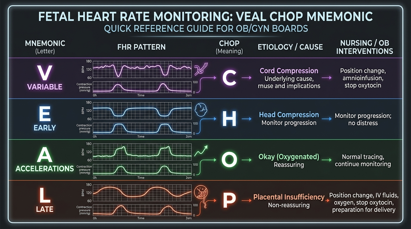
VEAL CHOP Fetal Heart Rate Monitoring Tracings
Cardiotocograph interpretation guide using the VEAL CHOP mnemonic: Variable = Cord compression; Early = Head compression; Accelerations = Okay (reassuring oxygenation); Late = Placental insufficiency. Recurrent late decelerations require immediate intrauterine resuscitation (maternal left lateral positioning, IV fluid bolus, 100% O2, stop oxytocin).
Deceleration Patterns & Pathophysiology (VEAL CHOP)
Clinical Matrix
Deceleration PatternMorphology & TimingEtiology / MechanismClinical Action
Variable Decelerations (V → C)Abrupt drop to nadir in < 30 sec, jagged shapeUmbilical Cord CompressionMaternal repositioning; Amnioinfusion for recurrent variables
Early Decelerations (E → H)Gradual symmetric drop matching contraction mirror-imageHead Compression (vagal reflex)Benign physiologic finding; no intervention needed, check dilation
Accelerations (A → O)Abrupt increase ≥ 15 bpm above baseline lasting ≥ 15 secOkay / Reassuring fetal oxygenationHallmark of fetal well-being and absence of acidosis
Late Decelerations (L → P)Gradual symmetric drop starting after contraction peakUteroplacental InsufficiencyIntrauterine resuscitation (stop pitocin, fluids, O2, lateral tilt); deliver if Category III
Board Trap — Shoulder Dystocia Emergency Sequence
High-Yield Board Trap & Alert
Shoulder dystocia occurs when the anterior fetal shoulder impacts behind the maternal pubic symphysis, signaled by the 'turtle sign' (fetal head retracting against perineum). Fundal pressure is STRICTLY CONTRAINDICATED because it impacts the shoulder harder and causes uterine rupture and brachial plexus injury (Erb-Duchenne palsy, C5–C6 'waiter's tip'). The correct initial actions are: Call for help, McRoberts maneuver (extreme hyperflexion and abduction of maternal hips onto abdomen), and simultaneous Suprapubic pressure directed downward and laterally to dislodge the shoulder. If unsuccessful, proceed to internal maneuvers (Rubin maneuver, Woods screw maneuver, delivery of posterior arm).

Chapter 3 Quick-Check Self-Assessment

Test your clinical reasoning before moving to the next chapter
3 Vignettes
Board Vignette #1 Single Best Answer
A 24-year-old G1P0 at 40 weeks is in labor. At 6 cm dilation, membranes are ruptured. An intrauterine pressure catheter is placed and confirms 230 Montevideo units of adequate uterine contractions. Four hours later, a sterile vaginal exam demonstrates no cervical progression (still 6 cm, 80% effaced, station 0). Fetal heart rate is Category I. What is the most appropriate next step in management?
Board Vignette #2 Single Best Answer
A 28-year-old G2P1 woman at 39 weeks gestation is in active labor. Continuous fetal heart rate monitoring displays recurrent, abrupt decreases in fetal heart rate from a baseline of 145 bpm down to 80 bpm. The onset of the deceleration to the nadir is 15 seconds. These decelerations occur inconsistently with uterine contractions and vary in duration and depth. What is the underlying pathophysiologic etiology?
Board Vignette #3 Single Best Answer
A 26-year-old delivers a 4,200 g infant via uncomplicated vaginal delivery. Ten minutes post-delivery, brisk continuous vaginal bleeding occurs. Physical examination reveals a soft, boggy, enlarged uterus palpable 3 cm above the umbilicus. Bimanual uterine massage and high-dose IV oxytocin infusion fail to stop the bleeding. The patient has a severe history of asthma requiring frequent nebulizers. What is the most appropriate next uterotonic agent?
Estimated study time: 13 min
Chapter 4 • Obstetric Complications & 3rd Trimester Bleeding
12 min TOC

Third-Trimester Vaginal Bleeding & Obstetric Catastrophes

Placenta Previa, Placental Abruption, Vasa Previa, and Uterine Rupture

Clinical Overview & Board Focus

Third-trimester bleeding requires immediate differentiation between maternal bleeding (previa, abruption, uterine rupture) and fetal bleeding (vasa previa). Digital cervical examination is strictly forbidden until placenta previa has been definitively excluded by transvaginal ultrasonography.

4.1 Third-Trimester Vaginal Bleeding Matrix

Placenta Previa occurs when the placenta implants over or immediately adjacent to the internal cervical os. It classically presents as painless, bright red vaginal bleeding in the second or third trimester. Risk factors include prior Cesarean deliveries, prior uterine curettage, multiparity, advanced maternal age, and multiple gestations. Transvaginal ultrasound (TVUS) is exceptionally safe and is the gold standard imaging modality. Digital pelvic examination is strictly contraindicated prior to ultrasound confirmation, as probing fingers can directly puncture the placental edge and provoke catastrophic hemorrhage. Complete placenta previa requires scheduled Cesarean delivery at 36 0/7 to 37 6/7 weeks.

Placental Abruption (abruptio placentae) is the premature separation of a normally implanted placenta from the uterine decidua prior to delivery. It presents classically as painful, dark vaginal bleeding accompanied by severe hypertonic uterine contractions, abdominal tenderness, and non-reassuring fetal heart rate tracings (recurrent late decelerations or fetal bradycardia). However, up to 20% of abruptions are 'concealed', in which hemorrhage is trapped entirely behind the placenta, producing severe pain and hypovolemic shock without external bleeding. Chronic hypertension, preeclampsia, cocaine use, tobacco use, and maternal abdominal trauma are premier risk factors. Retroplacental hematoma can release tissue factor into maternal circulation, precipitating severe Disseminated Intravascular Coagulation (DIC).

Vasa Previa occurs when aberrant fetal blood vessels course through the fetal membranes across the internal cervical os, unsupported by the placenta or umbilical cord (velamentous cord insertion or bilobed placenta). When amniotic membranes rupture (spontaneous or artificial amniotomy), these fetal vessels tear, producing painless vaginal bleeding accompanied by rapid, catastrophic fetal bradycardia or a sinusoidal fetal heart rate pattern. Bleeding is 100% FETAL blood; the Apt test or Kleihauer-Betke stain identifies fetal hemoglobin. Vasa previa mandates emergent Cesarean delivery within minutes to prevent total fetal exsanguination.

Differential Diagnosis of 3rd Trimester Vaginal Bleeding
Clinical Matrix
EtiologyPain StatusBleeding CharacterFetal Heart Rate StatusPrimary Management
Placenta PreviaPainlessBright red, variable amountUsually reassuring initiallyNo digital exam; TVUS confirmation; Cesarean at 36–37 wk
Placental AbruptionPainfulDark red (or concealed)Non-reassuring (late decels, bradycardia)Aggressive IV hydration, crossmatch blood, emergent delivery if unstable or fetal distress
Vasa PreviaPainlessBright red upon membrane ruptureRapid fetal bradycardia / sinusoidal patternEmergent STAT Cesarean section (fetal exsanguination imminent)
Uterine RuptureSevere tearing pain; contractions stopVaginal bleeding + intra-abdominal bloodCatastrophic bradycardia; loss of fetal stationEmergent laparotomy, Cesarean delivery, and uterine repair or hysterectomy
Board Trap — Uterine Rupture Hallmark
High-Yield Board Trap & Alert
Uterine rupture occurs almost exclusively in women undergoing a Trial of Labor After Cesarean (TOLAC) with a prior low transverse hysterotomy (risk 0.5–1%, and much higher with prior classical vertical incisions). The hallmark clinical sign is sudden loss of fetal station (the fetal presenting part, which was engaged at +1 or +2, suddenly retracts into the abdominal cavity on pelvic exam) accompanied by sudden, severe localized pain and cessation of previously regular uterine contractions on tocodynamometry. Emergent exploratory laparotomy is required immediately.

Chapter 4 Quick-Check Self-Assessment

Test your clinical reasoning before moving to the next chapter
3 Vignettes
Board Vignette #1 Single Best Answer
A 29-year-old G3P1 at 34 weeks presents to triage with sudden-onset bright red vaginal bleeding. She has no abdominal pain or contractions. Vitals: BP 118/74 mmHg, HR 80 bpm. Fetal heart rate baseline is 140 bpm with moderate variability. What clinical maneuver is strictly contraindicated at this time?
Board Vignette #2 Single Best Answer
A 31-year-old G3P2 female at 32 weeks gestation arrives at the labor and delivery unit reporting sudden-onset, painless, bright red vaginal bleeding that soaked two sanitary pads. She has had no uterine contractions and feels normal fetal movement. Vital signs are normal, and the abdomen is soft and non-tender. Fetal heart rate is 140 bpm with moderate variability. Which of the following procedures is strictly contraindicated?
Board Vignette #3 Single Best Answer
A 23-year-old primigravida has been pushing actively in the second stage of labor for 3.5 hours with an epidural. The fetal head is at +3 station, direct occiput anterior. Fetal heart rate baseline is 140 bpm with moderate variability. The maternal cervix is completely dilated and effaced. What is the diagnosis and next step?
Estimated study time: 12 min
Chapter 5 • Postpartum Care & Complications
13 min TOC

Postpartum Hemorrhage & Puerperal Infections

The 4 T's of PPH, Uterotonic Pharmacotherapy, Postpartum Endometritis, and Septic Pelvic Thrombophlebitis

Clinical Overview & Board Focus

Postpartum hemorrhage (PPH) is the leading cause of preventable maternal mortality worldwide. The emergency clinician and obstetrician must systematically evaluate the 4 T's (Tone, Trauma, Tissue, Thrombin) and implement rapid medical, mechanical, and surgical hemostasis.

5.1 Postpartum Hemorrhage & Stepwise Medical Resuscitation

Postpartum hemorrhage is defined as cumulative blood loss ≥ 1,000 mL or blood loss accompanied by signs or symptoms of hypovolemia within 24 hours of birth (regardless of delivery route). The etiologies are remembered as the '4 T's': Tone (Uterine Atony, accounting for 70–80% of cases), Trauma (cervical, vaginal, or perineal lacerations, uterine rupture), Tissue (retained placenta or succenturiate lobe), and Thrombin (coagulopathies). In uterine atony, the myometrium fails to contract down upon spiral arterioles, resulting in a soft, boggy, enlarged uterus above the umbilicus upon abdominal palpation.

Initial management of uterine atony begins with vigorous bimanual uterine massage and immediate administration of IV Oxytocin (Pitocin, 10–40 units in 1 L crystalloid). Bladder catheterization is performed to decompress the bladder, which mechanically impedes uterine involution. If atony persists, secondary uterotonic agents are deployed systematically: (1) Methylergonovine (Methergine, 0.2 mg IM; smooth muscle ergot alkaloid; strictly contraindicated in hypertension/preeclampsia due to profound peripheral vasoconstriction); (2) Carboprost tromethamine (Hemabate, 250 mcg IM; prostaglandin F2-alpha; strictly contraindicated in asthma due to severe bronchospasm); and (3) Misoprostol (Cytotec, 800–1,000 mcg PR or sublingual; prostaglandin E1 analogue).

If pharmacotherapy fails, mechanical intrauterine tamponade is initiated using a Bakri balloon. Refractory hemorrhage mandates immediate transition to operative intervention: exploratory laparotomy with uterine compression sutures (B-Lynch technique), bilateral hypogastric / internal iliac artery ligation (reducing pelvic pulse pressure), or definitive emergent peripartum hysterectomy. Massive transfusion protocols (1:1:1 ratio) and TXA (1 g IV) are administered simultaneously.

Uterotonic Pharmacotherapy Contraindication Matrix
Clinical Matrix
MedicationMechanismRoute & DosageAbsolute Board Contraindication
Oxytocin (Pitocin)Binds myometrial oxytocin receptors; rhythmic contractionsIV infusion (10–40 U in 1L NS/LR)None (first-line universal agent)
Methylergonovine (Methergine)Ergot alkaloid; sustained tetanic uterine contraction0.2 mg IM (never IV due to stroke risk)Hypertension, Preeclampsia, CAD (severe vasospasm)
Carboprost (Hemabate)Prostaglandin F2-alpha analogue250 mcg IM every 15–90 min (max 8 doses)Asthma / Bronchospasm (causes potent bronchoconstriction)
Misoprostol (Cytotec)Prostaglandin E1 analogue; cervical ripening and contraction800–1000 mcg sublingual or per rectumKnown hypersensitivity to prostaglandins
COMLEX Clinical Integration — Postpartum Sacral Dysfunction & Pelvic Diaphragm Rehabilitation
COMLEX & OPP Board Integration
Passage of the fetal head through the birth canal exerts massive mechanical forces upon the coccyx, pubic symphysis, and pelvic floor levator ani musculature. Postpartum examination frequently reveals unilateral sacral shear (sacral subluxation), bilateral sacral flexion, or pubic shear dysfunctions, manifesting as severe groin pain and difficulty ambulating. Furthermore, hypertonicity of the obturator internus and pubococcygeus impairs venous return from the pelvic plexus, promoting vulvar edema and hemorrhoids. Gentle counterstrain to pelvic tenderpoints, muscle energy for pubic shear, and CV4 cranial treatment accelerate maternal recovery and normalize autonomic outflow.

Chapter 5 Quick-Check Self-Assessment

Test your clinical reasoning before moving to the next chapter
3 Vignettes
Board Vignette #1 Single Best Answer
A 28-year-old G1P1 with preeclampsia with severe features undergoes normal spontaneous vaginal delivery. Ten minutes post-delivery, she has profuse vaginal bleeding (800 mL) and her uterus is soft, boggy, and palpated 3 cm above the umbilicus. Bimanual massage and IV oxytocin fail to contract the uterus. Blood pressure is 162/104 mmHg. Which secondary uterotonic is strictly contraindicated?
Board Vignette #2 Single Best Answer
A 25-year-old G1P1 woman develops a temperature of 38.9°C (102.0°F), chills, and lower abdominal pain on postpartum day 2 following an urgent cesarean delivery for arrested labor. Examination reveals purulent, foul-smelling lochia and marked exquisite uterine fundal tenderness. WBC count is 22,000/µL. What is the most appropriate first-line antimicrobial regimen?
Board Vignette #3 Single Best Answer
A 22-year-old female presents with lower abdominal pain, purulent cervical discharge, and severe cervical motion tenderness ('chandelier sign'). Urine pregnancy test is negative. What is the most appropriate outpatient antimicrobial regimen?
Estimated study time: 13 min
Chapter 6 • Benign Gynecology, Pelvic Pain & AUB
13 min TOC

Benign Gynecology, Abnormal Uterine Bleeding & Pelvic Pain

PALM-COEIN Classification, Endometriosis, Adenomyosis, Uterine Leiomyomas, and Ovarian Torsion

Clinical Overview & Board Focus

Abnormal uterine bleeding (AUB) and chronic pelvic pain are ubiquitous clinical presentations. The International Federation of Gynecology and Obstetrics (FIGO) PALM-COEIN system organizes bleeding into structural vs. non-structural causes. Ovarian torsion represents a time-sensitive surgical emergency requiring immediate diagnostic laparoscopy.

6.1 PALM-COEIN Classification & Differential Diagnosis of Chronic Pelvic Pain

Abnormal Uterine Bleeding in non-pregnant reproductive-aged women is categorized into Structural etiologies (PALM: Polyp, Adenomyosis, Leiomyoma, Malignancy/hyperplasia) and Non-structural etiologies (COEIN: Coagulopathy, Ovulatory dysfunction, Endometrial, Iatrogenic, Not otherwise classified). In any woman over age 45 (or < 45 with risk factors like unopposed estrogen, obesity, PCOS, or failure to respond to medical therapy), endometrial biopsy is MANDATORY to rule out endometrial hyperplasia and carcinoma.

Endometriosis is defined as the presence of functional endometrial glands and stroma outside the uterine cavity, most commonly involving the ovaries (forming 'chocolate cysts' or endometriomas), uterosacral ligaments, and pouch of Douglas. It classically presents with the triad of Dysmenorrhea (painful menstruation), Dyspareunia (deep dyspareunia), and Dyschezia (pain with defecation during menses), often accompanied by subfertility. Physical examination may reveal a fixed, retroverted uterus and nodularity along the uterosacral ligaments. Definitive diagnosis requires laparoscopic visualization with histologic biopsy. First-line medical therapy involves combined oral contraceptives (COCs) or progestins; second-line therapy includes GnRH receptor agonists (Leuprolide) or GnRH antagonists (Elagolix) with add-back progestin therapy.

Adenomyosis occurs when endometrial basal glands and stroma invade the myometrium, causing reactive myometrial hypertrophy. It classically presents in parous women in their late 30s or 40s with heavy menstrual bleeding and severe dysmenorrhea. On physical exam, the uterus is symmetrically enlarged, boggy, and tender (distinguished from leiomyomas, which produce an asymmetrically enlarged, firm, non-tender multinodular uterus). Pelvic ultrasound or MRI reveals myometrial heterogeneity and junctional zone thickening > 12 mm. Definitive cure is hysterectomy; levonorgestrel-releasing IUD provides excellent medical symptom relief.

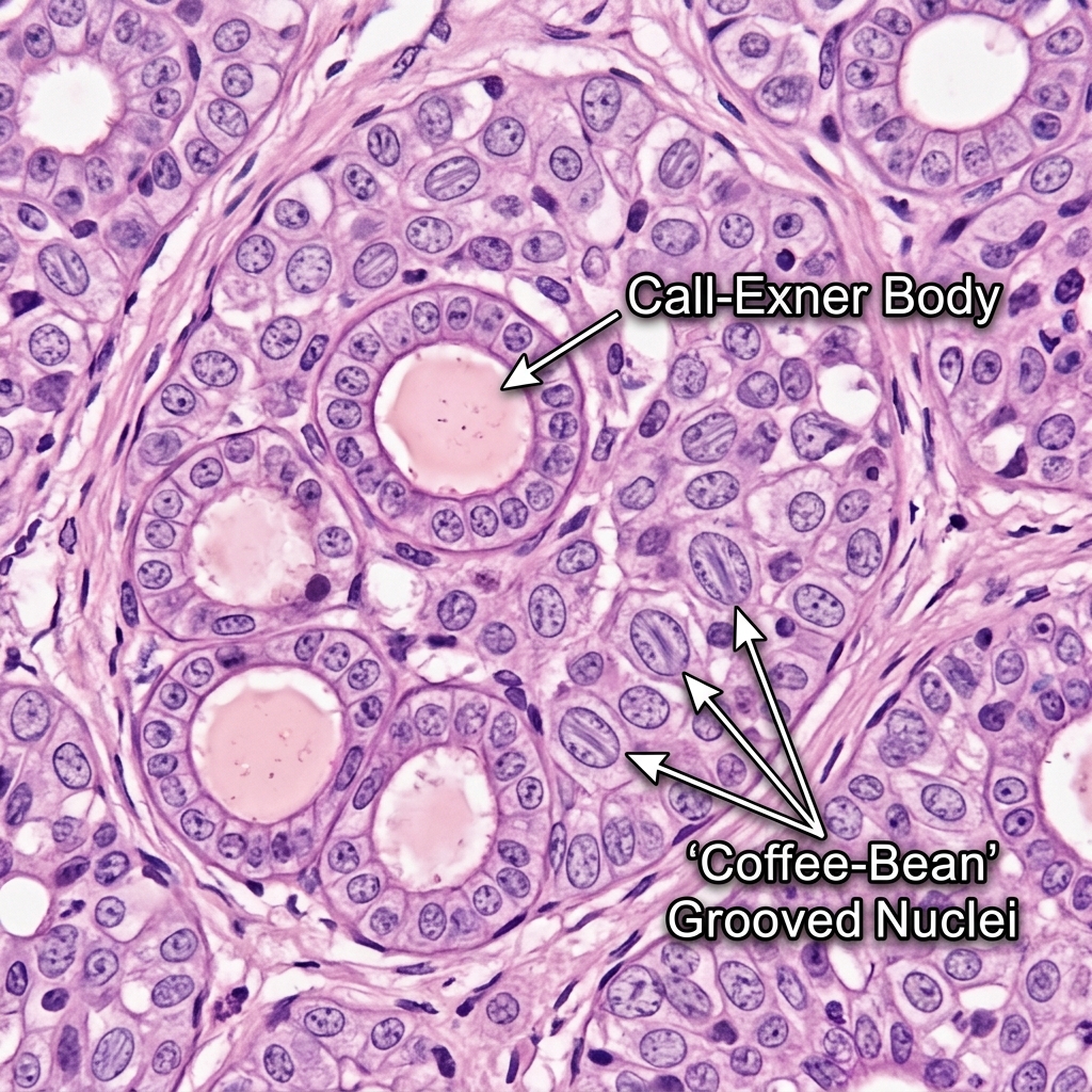
Granulosa Cell Tumor Call-Exner Bodies
Histopathology of a Granulosa Cell Tumor of the ovary demonstrating microfollicular clusters of granulosa cells surrounding eosinophilic fluid spaces resembling immature follicles (Call-Exner bodies), alongside cells with 'coffee-bean' grooved nuclei. These tumors secrete estrogen (causing endometrial hyperplasia / postmenopausal bleeding) and inhibin (used as a tumor marker).
Pelvic Pain & Uterine Pathology Comparison
Clinical Matrix
ConditionPhysical Exam HallmarkDiagnostic Imaging FindingFirst-Line Treatment
EndometriosisFixed, retroverted uterus, nodularity of uterosacral ligamentsGround-glass appearance on TVUS (endometrioma)Combined OCPs or Progestins; Laparoscopic ablation
AdenomyosisGlobular, symmetrically enlarged, 'boggy', tender uterusJunctional zone thickening > 12 mm on MRI/TVUSLNG-IUD or OCPs; definitive cure is Hysterectomy
Uterine Leiomyoma (Fibroids)Asymmetrically enlarged, firm, non-tender, irregular contoured uterusWell-circumscribed hypoechoic pelvic masses on TVUSCOCs, LNG-IUD, Tranexamic acid, Myomectomy (preserves fertility), Hysterectomy
Ovarian TorsionSudden-onset severe unilateral lower quadrant pain, nausea/vomitingEnlarged, edematous ovary with absent or diminished Doppler flowEmergent Laparoscopy with detorsion and ovarian preservation
Board Trap — Doppler Flow in Ovarian Torsion
High-Yield Board Trap & Alert
The presence of arterial Doppler flow on pelvic ultrasound does NOT rule out ovarian torsion! Because the ovary has a dual blood supply (the ovarian artery from the abdominal aorta and the ovarian branch of the uterine artery), and because venous and lymphatic outflow are occluded long before arterial inflow is completely compromised, up to 30% of surgically proven ovarian torsions demonstrate normal arterial Doppler signals on ultrasound. If a patient presents with sudden-onset severe sharp unilateral lower quadrant pain, tender adnexal mass, and vomiting, proceed to emergent diagnostic laparoscopy regardless of Doppler report.

Chapter 6 Quick-Check Self-Assessment

Test your clinical reasoning before moving to the next chapter

3 Vignettes
Board Vignette #1 Single Best Answer
A 42-year-old multiparous female presents with progressively worsening heavy menstrual bleeding and severe dysmenorrhea. Bimanual exam reveals a symmetrically enlarged, globular, tender, and boggy uterus. Transvaginal ultrasound demonstrates myometrial heterogeneity and diffuse junctional zone thickening to 16 mm. What is the definitive diagnosis?
Board Vignette #2 Single Best Answer
A 27-year-old sexually active woman presents with 6 weeks of amenorrhea followed by acute right lower quadrant abdominal pain and light vaginal spotting. Serum beta-hCG is 2,400 mIU/mL. Transvaginal ultrasonography demonstrates an empty uterine cavity with an endometrial thickness of 8 mm, a 2.5-cm complex right adnexal mass with a 'ring-of-fire' vascular pattern, and no free fluid in the cul-de-sac. Her vital signs are stable. Which of the following is the most appropriate initial management?
Board Vignette #3 Single Best Answer
A 29-year-old female presents with severe cyclic pelvic pain, deep dyspareunia, and dyschezia that worsens during menses. Pelvic examination reveals tenderness along the uterosacral ligaments with fixed retroversion of the uterus. What is the definitive gold-standard diagnostic modality?
Estimated study time: 13 min
Chapter 7 • Gynecologic Oncology & Cervical Pathology
13 min TOC

Gynecologic Malignancies & Cervical Cancer Screening

Cervical Dysplasia Management, Endometrial Carcinoma, Epithelial Ovarian Cancer, and Gestational Trophoblastic Disease

Clinical Overview & Board Focus

Gynecologic oncology covers cervical screening guidelines (USPSTF/ASCCP), workup of postmenopausal bleeding, identification of ovarian masses, and risk factors associated with hereditary cancer syndromes (BRCA1/2, Lynch syndrome).

7.1 Cervical Dysplasia Guidelines & Endometrial/Ovarian Carcinoma

Cervical cancer screening guidelines are strictly tested: Screening begins at age 21 regardless of sexual debut. Women age 21–29 undergo cervical cytology (Pap smear) alone every 3 years (HPV co-testing is NOT recommended under age 30 due to high transient HPV clearance rates). For women age 30–65, three acceptable options exist: (1) Cytology alone every 3 years; (2) High-risk HPV (hrHPV) testing alone every 5 years; or (3) Co-testing (Cytology + hrHPV) every 5 years. Screening discontinues at age 65 if adequate prior negative screening is documented (3 consecutive negative cytology or 2 negative co-tests in past 10 years, with the most recent test within 5 years) and no history of CIN 2+ within 25 years.

Management of abnormal Pap smears follows ASCCP risk-based guidelines. In women ≥ 25 years with Atypical Squamous Cells of Undetermined Significance (ASC-US), reflex HPV testing is performed: if HPV-negative, repeat cytology in 3 years; if HPV-positive, proceed directly to Colposcopy. In patients with High-Grade Squamous Intraepithelial Lesions (HSIL) or Atypical Glandular Cells (AGC), immediate Colposcopy (with endocervical curettage [ECC] and endometrial biopsy for AGC in women ≥ 35) is indicated. During colposcopy, application of 3–5% acetic acid reveals acetowhite epithelium and abnormal vascular punctation or mosaicism, guiding directed punch biopsies.

Postmenopausal Bleeding (PMB) is considered Endometrial Carcinoma until proven otherwise. Endometrial cancer is the most common gynecologic malignancy in the United States, driven by unopposed estrogen exposure (obesity, nulliparity, late menopause, tamoxifen therapy, and Lynch syndrome / HNPCC). Initial evaluation requires either Transvaginal Ultrasound (evaluating endometrial stripe thickness; thickness ≤ 4 mm safely excludes cancer with 99% NPV) or definitive Endometrial Biopsy (mandatory if stripe > 4 mm or bleeding recurs).

Epithelial Ovarian Carcinoma typically presents insidiously in postmenopausal women with non-specific abdominal bloating, early satiety, pelvic fullness, and ascites. Physical exam reveals a fixed, solid, nodular adnexal mass. Serum CA-125 is elevated, but is non-specific (can be elevated in endometriosis, fibroids, cirrhosis, and pelvic inflammatory disease); its primary utility lies in monitoring response to surgical debulking and platinum-based chemotherapy. Any postmenopausal woman with a complex, solid adnexal mass and elevated CA-125 warrants immediate referral to Gynecologic Oncology for exploratory staging laparotomy.

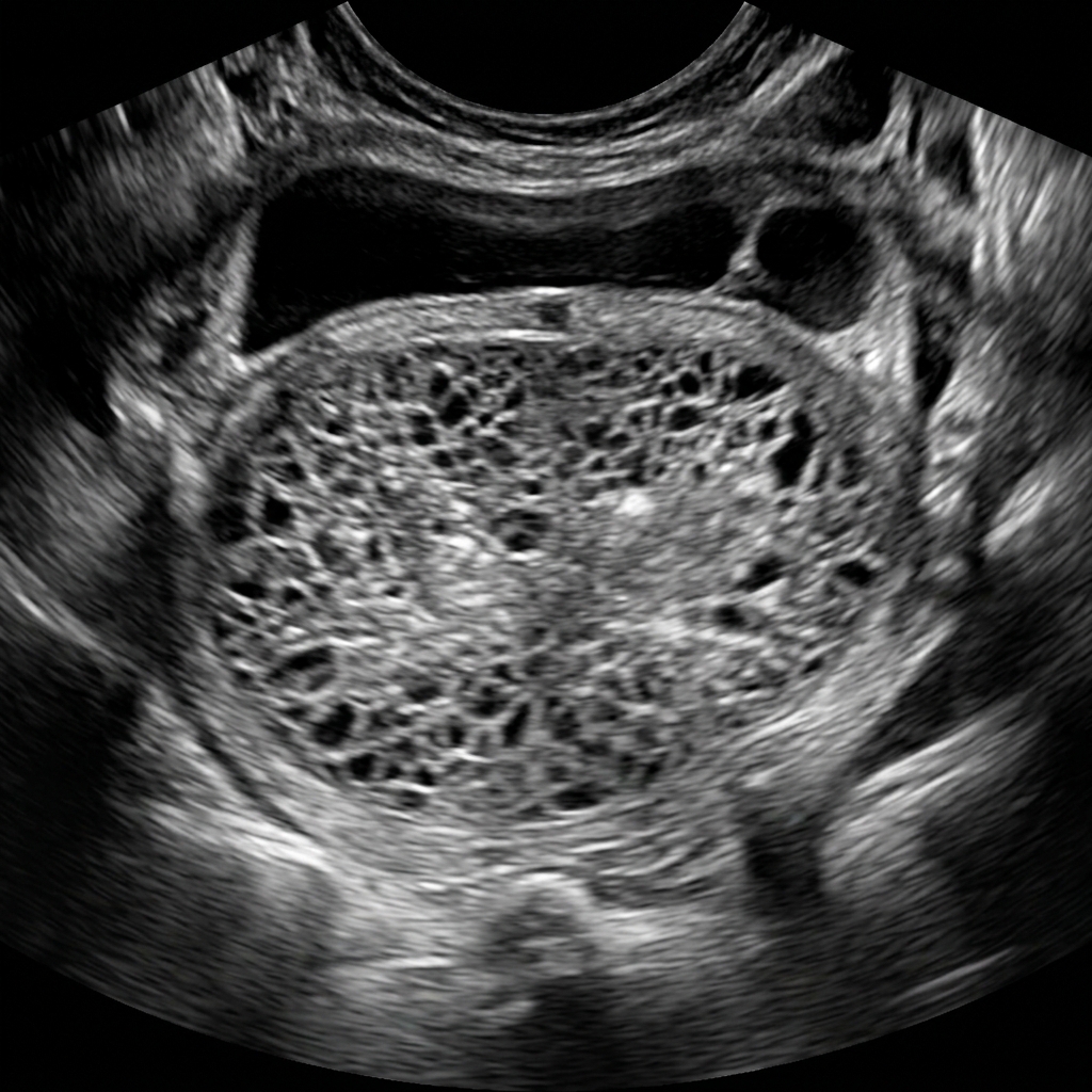
Snowstorm appearance on ultrasound in complete mole
Transvaginal ultrasound of Complete Hydatidiform Mole, demonstrating the pathognomonic 'snowstorm' or 'Swiss-cheese' appearance consisting of a complex hyperechoic central intrauterine mass containing innumerable microcystic vesicles (hydropic trophoblastic chorionic villi) without fetal tissue or amniotic sac. Complete mole arises from paternal duplication or dispermy in an empty ovum (46,XX or 46,XY) and presents with markedly elevated beta-hCG (>100,000 mIU/mL).
High-Yield Clinical Takeaway — Gestational Trophoblastic Disease
High-Yield Clinical Takeaway
Complete Hydatidiform Mole results from fertilization of an empty enucleated ovum by one sperm that duplicates (46,XX, 90%) or two sperm (46,XY). It presents with markedly elevated beta-hCG (> 100,000 mIU/mL), hyperemesis gravidarum, bilateral theca lutein ovarian cysts, uterine size larger than gestational age, and early preeclampsia (< 20 weeks). Pelvic ultrasound demonstrates the classic 'snowstorm' or 'swiss cheese' vesicular pattern without fetal parts. Treatment is immediate suction curettage followed by serial weekly quantitative beta-hCG monitoring until zero for 6 months (with strict contraception) to monitor for Gestational Choriocarcinoma.

Chapter 7 Quick-Check Self-Assessment

Test your clinical reasoning before moving to the next chapter

3 Vignettes
Board Vignette #1 Single Best Answer
A 58-year-old postmenopausal female presents with two episodes of light vaginal spotting. Her last menstrual period was 7 years ago. Pelvic exam is unremarkable. What is the mandatory next diagnostic step in this patient?
Board Vignette #2 Single Best Answer
A 61-year-old postmenopausal woman presents with light vaginal bleeding for the past 3 weeks. She completed menopause at age 52 and has not had bleeding in 9 years. Her BMI is 34 kg/m², and medical history is significant for hypertension and type 2 diabetes. Pelvic exam is unremarkable. Transvaginal ultrasound demonstrates an endometrial stripe measuring 8 mm. Which of the following is the most appropriate next step in management?
Board Vignette #3 Single Best Answer
A 25-year-old female presents with irregular menses every 45–60 days, severe facial hirsutism, and BMI 34 kg/m2. Total testosterone is mildly elevated and pelvic ultrasound reveals bilateral enlarged ovaries with >20 subcapsular follicles ('string of pearls'). She desires pregnancy. What is the first-line pharmacologic ovulation induction agent?
Estimated study time: 13 min
Chapter 8 • Reproductive Endocrinology, Infertility & Contraception
13 min TOC

Reproductive Endocrinology, Amenorrhea & Contraception

Primary and Secondary Amenorrhea Algorithms, PCOS, Infertility Workup, and Contraceptive Selection

Clinical Overview & Board Focus

Reproductive endocrinology tests systematic hormone feedback loops involving the hypothalamus, anterior pituitary, and ovaries. Understanding the diagnostic algorithms for primary vs. secondary amenorrhea and contraceptive efficacy and contraindications is high-yield for board success.

8.1 Primary and Secondary Amenorrhea Diagnostic Algorithms

Primary Amenorrhea is defined as the absence of menarche by age 13 in the absence of secondary sexual characteristics (breast development), or by age 15 in the presence of normal secondary sexual characteristics. Initial evaluation begins with physical examination (assessing breast development, indicating estrogen exposure) and Pelvic Ultrasound (confirming presence or absence of a uterus). If the uterus is absent, obtain a karyotype to differentiate Müllerian Agenesis (Mayer-Rokitansky-Küster-Hauser syndrome: 46,XX with normal ovaries, normal female testosterone levels, normal axillary/pubic hair) from Androgen Insensitivity Syndrome (AIS: 46,XY with male testosterone levels, absent pubic/axillary hair, and cryptorchid intra-abdominal testes requiring gonadectomy after puberty to prevent dysgerminoma/gonadoblastoma).

If the uterus is present but breasts are absent (lack of estrogen), obtain serum FSH and LH. Elevated FSH indicates Hypergonadotropic Hypogonadism (primary ovarian failure, most commonly Turner Syndrome: 45,XO, streak ovaries, short stature, webbed neck, bicuspid aortic valve, coarctation of the aorta). Low or normal FSH indicates Hypogonadotropic Hypogonadism (hypothalamic or pituitary failure, such as Kallmann Syndrome: failure of GnRH neural migration from olfactory placode, characterized by anosmia and absent puberty).

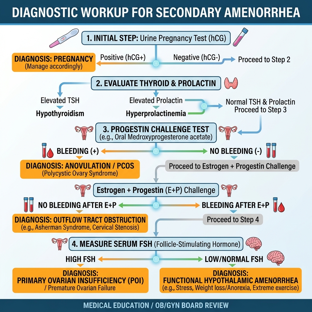
Secondary Amenorrhea is defined as the cessation of menses for ≥ 3 months in women with previously regular cycles, or ≥ 6 months in women with oligomenorrhea. The initial and most critical step is ALWAYS a urine pregnancy test (beta-hCG). Once pregnancy is excluded, check serum TSH (hypothyroidism increases TRH, stimulating prolactin release), Prolactin (prolactinoma or dopamine antagonist drugs), and FSH. The Progestin Challenge Test (oral Medroxyprogesterone acetate 10 mg × 10 days) assesses endogenous estrogen status: withdrawal bleeding indicates anovulation with intact estrogen production (most commonly Polycystic Ovary Syndrome [PCOS]); absence of withdrawal bleeding indicates either severe hypoestrogenism (hypothalamic amenorrhea, premature ovarian insufficiency) or an outflow tract obstruction (Asherman syndrome from prior intrauterine curettage).

Secondary Amenorrhea Diagnostic Workup Algorithm
Stepwise diagnostic flowchart for Secondary Amenorrhea: Rule out pregnancy first with urine/serum beta-hCG, followed by serum TSH and Prolactin. If negative, perform the Progestin Challenge Test (oral medroxyprogesterone × 10 days). Bleeding confirms anovulation/PCOS (unopposed estrogen). Absence of withdrawal bleeding prompts Estrogen + Progestin priming: withdrawal bleeding indicates hypothalamic-pituitary or ovarian failure (differentiated by FSH/LH levels), whereas failure to bleed establishes an outflow tract obstruction (Asherman syndrome or cervical stenosis).
Contraceptive Methods & High-Yield Contraindications
Clinical Matrix
Contraceptive ModalityMechanism of ActionTypical Failure RateKey Contraindications / Board Traps
Combined Hormonal (Pill, Patch, Ring)Suppresses LH/FSH surge (inhibits ovulation) + thickens cervical mucus7–9% (typical)Age ≥ 35 smoking ≥ 15 cig/day, migraine with aura, uncontrolled HTN (> 160/100), history of DVT/PE/stroke, breast cancer
Levonorgestrel IUD (Mirena/Kyleena)Thickens cervical mucus, causes endometrial atrophy, reduces menses0.2%Active pelvic infection (PID within 3 months), unexplained uterine bleeding, uterine cavity distortion
Copper IUD (ParaGard)Sterile inflammatory response toxic to sperm and ova; non-hormonal0.8%Wilson disease, active pelvic infection, severe dysmenorrhea/menorrhagia (increases bleeding)
Progestin Implant (Nexplanon)Etonogestrel rod; suppresses ovulation × 3–5 years0.05%Active breast cancer, severe hepatic disease; highly effective, causes irregular bleeding
Depot Medroxyprogesterone (DMPA)IM injection every 12 weeks; inhibits LH surge4%Long-term use (> 2 years) causes reversible loss of bone mineral density (BMD)
Board Trap — Migraine with Aura and Estrogen Contraindication
High-Yield Board Trap & Alert
A 28-year-old woman requests contraception. She reports unilateral throbbing headaches preceded by flashing zig-zag lights (scintillating scotoma) lasting 30 minutes. Combined estrogen-progestin oral contraceptives are ABSOLUTELY CONTRAINDICATED because estrogen increases ischemic stroke risk up to 7-fold in patients with migraine with aura! Recommend a progestin-only method (levonorgestrel IUD, progestin implant, progestin-only pill) or non-hormonal copper IUD.

Chapter 8 Quick-Check Self-Assessment

Test your clinical reasoning before moving to the next chapter

3 Vignettes
Board Vignette #1 Single Best Answer
A 16-year-old female presents with primary amenorrhea. Physical exam reveals Tanner stage 4 breast development but completely absent axillary and pubic hair. Pelvic ultrasound reveals a blind-ending vaginal pouch and absent uterus and ovaries. Bilateral masses are palpated in the labia majora. Serum testosterone is in the adult male range. What is the diagnosis and karyotype?
Board Vignette #2 Single Best Answer
A 24-year-old nulligravid female presents with irregular menses every 60 to 90 days, severe cystic acne, and coarse facial hair on her chin and upper lip. Her BMI is 31 kg/m². Pelvic ultrasonography reveals multiple small subcapsular ovarian cysts in a 'string-of-pearls' distribution. She desires pregnancy. In addition to lifestyle modification and weight loss, which of the following is the first-line medication to induce ovulation?
Board Vignette #3 Single Best Answer
A 61-year-old postmenopausal female presents with painless vaginal spotting for 3 weeks. Her last menstrual period was 10 years ago. Pelvic ultrasound reveals a thickened endometrial stripe measuring 9 mm. What is the next mandatory diagnostic step?
Estimated study time: 13 min
Chapter 9 • Infectious Diseases, STIs & Vaginitis
13 min TOC

Gynecologic Infectious Diseases, Vaginitis & STIs

Vaginitis Triad, Pelvic Inflammatory Disease, Fitz-Hugh-Curtis Syndrome, and Genital Ulcer Differentials

Clinical Overview & Board Focus

Infectious diseases of the female reproductive tract range from common superficial vaginitis to life-threatening tubo-ovarian abscesses and ascending upper genital tract scarring that produces ectopic pregnancy and tubal factor infertility.

9.1 The Vaginitis Triad, PID & Genital Ulcerative Diseases

Vaginitis is evaluated via physical exam, vaginal pH, 10% KOH whiff test, and wet mount microscopy. Bacterial Vaginosis (BV) is an overgrowth of anaerobic organisms (Gardnerella vaginalis) replacing hydrogen peroxide-producing Lactobacillus. It presents with thin, off-white/gray homogenous discharge with an amine 'fishy' odor. Diagnostic criteria (Amsel criteria, ≥ 3 of 4): vaginal pH > 4.5, positive whiff test on KOH, thin homogenous discharge, and clue cells (> 20% vaginal epithelial cells coated in bacteria) on saline wet mount. Treatment is oral or intravaginal Metronidazole × 7 days; sexual partners do NOT require treatment.

Trichomoniasis (Trichomonas vaginalis) is a sexually transmitted protozoan infection presenting with profuse, frothy, yellow-green malodorous discharge, vulvar erythema, and punctate cervical hemorrhages ('strawberry cervix'). Vaginal pH is > 4.5. Wet mount microscopy reveals motile flagellated trichomonads. Treatment is Metronidazole (500 mg BID × 7 days or 2 g single dose); sexual partners MUST be treated simultaneously, and patients must avoid alcohol (disulfiram-like reaction).

Candida Vulvovaginitis (Candida albicans) presents with thick, white, clumpy 'cottage-cheese' discharge, severe pruritus, and labial erythema. Vaginal pH is strictly NORMAL (4.0–4.5). KOH wet mount reveals pseudohyphae and budding yeast. Treatment is oral single-dose Fluconazole (150 mg PO) or topical azoles (Clotrimazole, Miconazole).

Pelvic Inflammatory Disease (PID) is an ascending polymicrobial infection originating from Chlamydia trachomatis, Neisseria gonorrhoeae, or enteric anaerobes. Minimum clinical diagnostic criteria include pelvic/lower abdominal pain plus at least one: cervical motion tenderness ('chandelier sign'), uterine tenderness, or adnexal tenderness. Perihepatic capsular inflammation produces right upper quadrant pain and 'violin-string' adhesions (Fitz-Hugh-Curtis syndrome). Outpatient treatment: Ceftriaxone (500 mg IM) + Doxycycline (100 mg BID × 14 days) + Metronidazole (500 mg BID × 14 days). Inpatient criteria (pregnancy, failed outpatient therapy, pelvic peritonitis, tubo-ovarian abscess [TOA], inability to tolerate oral meds) require IV Cefotetan or Cefoxitin plus IV Doxycycline, or IV Ampicillin-Sulbactam plus Doxycycline.

Genital Ulcer Differential Diagnosis
Clinical Matrix
DiseaseCausative OrganismUlcer Morphology & PainLymphadenopathy Finding
Genital Herpes (HSV-1 / HSV-2)Herpes simplex virusMultiple painful, vesicular erosions with erythematous baseTender bilateral inguinal lymphadenopathy
Primary SyphilisTreponema pallidumSingle, painless chancre with clean base and indurated marginsPainless, rubbery bilateral inguinal lymphadenopathy
ChancroidHaemophilus ducreyiPainful, deep ulcer with ragged undermined borders and purulent basePainful, fluctuant suppurative inguinal lymphadenitis (bubo)
Lymphogranuloma Venereum (LGV)Chlamydia trachomatis (L1–L3)Small, transient, painless papule/ulcer that quickly heals unnoticedPainful, prominent inguinal lymphadenopathy with 'groove sign'
COMLEX Clinical Integration — Chapman's Reflexes in Pelvic Inflammatory Disease
COMLEX & OPP Board Integration
Pelvic inflammatory disease and adnexal pathology produce distinct viscerosomatic reflexes: Uterine tissue facilitation concentrates paraspinally at L1–L2, while the Ovaries and Fallopian Tubes project sympathetics to T10–T11 (lesser splanchnic nerve, superior mesenteric ganglion). The anterior Chapman's reflex point for the Uterus is located on the superior pubic ramus near the symphysis; the anterior Chapman's point for the Ovaries and Salpinges is located on the superior aspect of the pubic bone near the pubic tubercle. In severe pelvic infection, HVLA is contraindicated; gentle rib raising, thoracolumbar paraspinal release, and thoracic inlet opening reduce sympathetic tone and facilitate lymphatic drainage of the pelvic basin.

Chapter 9 Quick-Check Self-Assessment

Test your clinical reasoning before moving to the next chapter

3 Vignettes
Board Vignette #1 Single Best Answer
A 22-year-old female presents with intense vulvar itching and thick white vaginal discharge. Pelvic examination reveals labial erythema and thick adherent discharge resembling cottage cheese. Cervix is normal. Vaginal pH is 4.2. What microscopic finding confirms the diagnosis?
Board Vignette #2 Single Best Answer
A 26-year-old female presents with copious, malodorous, frothy yellow-green vaginal discharge and severe vulvar pruritus. Speculum examination reveals diffuse vaginal erythema with punctate erythematous hemorrhages on the cervix ('strawberry cervix'). Wet mount microscopy shows numerous motile, flagellated, pear-shaped organisms. What is the most appropriate management?
Board Vignette #3 Single Best Answer
A 54-year-old female presents with involuntary urine leakage during coughing, sneezing, and laughing. She denies urinary urgency or nocturia. Physical exam demonstrates cystocele and a positive cough stress test. What is the most appropriate first-line conservative management?
Estimated study time: 13 min