Skip to content
NBOME Internal Medicine COMAT & Level 2-CE NBME Internal Medicine Shelf Exam & Step 2 CK

Internal Medicine COMAT Command Center Internal Medicine Shelf Exam Command Center

Comprehensive, high-yield clinical command center for the NBOME Internal Medicine COMAT shelf and COMLEX Level 2-CE. Ingests 4,400+ IM clerkship cards across cardiology, pulmonology, gastroenterology, nephrology, endocrinology, infectious disease, and osteopathic inpatient medicine.

72 Cards Extracted 3 High-Yield Protocols 4 Osteopathic Rules 4 High-Yield Pearls
Section 01 • Osteopathic Principles

"Don't Miss" COMAT Osteopathic Pearls

High-frequency somatic dysfunctions, viscerosomatics, autonomic reflexes, and treatment rules

Tested heavily on NBOME Shelf

Cardiac Viscerosomatics & Autonomics

Sympathetics: T1–T5 via cervical and upper thoracic ganglia (Left side = left ventricle and predisposes to ventricular arrhythmias/infarction; Right side = SA node and supraventricular tachycardias). Parasympathetics: Vagus nerve (CN X). Anterior Chapman: 2nd ICS lateral to sternum. Posterior Chapman: Between T2–T3 spinous processes.

Pulmonary Autonomics & Rib Mechanics

Sympathetics: T2–T7. Parasympathetics: Vagus nerve (CN X). Anterior Chapman: 3rd & 4th ICS adjacent to sternum. Inpatient OPP: Rib raising normalizes sympathetic tone and reduces airway hypersensitivity. Treating exhalation somatic dysfunctions (1st rib down) elevates the chest cage and improves functional residual capacity in COPD/pneumonia.

Gastrointestinal Ganglia Rules

Celiac Ganglion (T5–T9): Stomach, liver, gallbladder, spleen, portions of pancreas, 1st/2nd duodenum. Superior Mesenteric Ganglion (T10–T11): 3rd/4th duodenum, jejunum, ileum, ascending colon, proximal 2/3 transverse colon. Inferior Mesenteric Ganglion (T12–L2): Distal 1/3 transverse colon, descending colon, sigmoid colon, rectum. Parasympathetics: Vagus for Celiac & SMG; Pelvic splanchnics (S2–S4) for IMG.

Renal & Adrenal Autonomics

Kidneys: T10–T11 sympathetics; Vagus parasympathetics. Ureters: T10–T11 (upper) and T12–L2 (lower). Anterior Chapman: 1 inch superior and 1 inch lateral to umbilicus. Adrenals: T8–T10 sympathetics (innervate medulla directly via preganglionic fibers). Anterior Chapman: 2 inches superior and 1 inch lateral to umbilicus.

Section 01 • Clinical Foundations

"Don't Miss" Internal Medicine Clinical Pearls & Shelf Traps

High-frequency diagnostic pitfalls, gold-standard criteria, and next-best-step clinical rules

Tested heavily on NBME Shelf & Step 2 CK

Heart Failure GDMT Quadruple Therapy & Mortality Reduction

HFrEF (LVEF <= 40%) mortality reduction requires four pillar classes: 1. ARNI (Sacubitril/Valsartan preferred over ACEi/ARB; mandatory 36-hour washout when switching from ACEi to avoid angioedema); 2. Evidence-based Beta-blocker (Carvedilol, Metoprolol succinate, or Bisoprolol—titrate only when euvolemic); 3. Mineralocorticoid Receptor Antagonist (Spironolactone or Eplerenone—monitor K+ and renal function); 4. SGLT2 inhibitor (Dapagliflozin or Empagliflozin—reduces HF hospitalizations regardless of diabetes status).

Hyponatremia Diagnostic Algorithm & Correction Traps

Step 1: Check serum osmolality (normal 275-295 -> pseudohyponatremia from hyperproteinemia/hyperlipidemia; high > 295 -> hyperglycemia [corrected Na = measured Na + 1.6 * (glucose - 100) / 100]; low < 275 -> true hypotonic hyponatremia). Step 2: Assess volume status (Hypovolemic -> check urine Na; Euvolemic -> SIADH [urine Osm > 100, urine Na > 40]; Hypervolemic -> HF, cirrhosis, nephrotic syndrome). Critical safety limit: Never correct chronic hyponatremia > 8 mEq/L in 24 hours to prevent irreversible Osmotic Demyelination Syndrome (central pontine myelinolysis).

Cirrhosis Decompensation: SBP, Varices & Hepatorenal Syndrome

Spontaneous Bacterial Peritonitis (SBP): Diagnostic paracentesis is mandatory in any acute decompensation; PMN count >= 250/mm³ confirms diagnosis. Treat immediately with IV 3rd-generation cephalosporin (Ceftriaxone or Cefotaxime) plus IV albumin (1.5 g/kg on day 1, 1.0 g/kg on day 3) to prevent hepatorenal syndrome. Acute Variceal Bleed: Airway protection, two large-bore IVs, IV Octreotide bolus + infusion, prophylactic IV Ceftriaxone, restrictive transfusion (target Hb 7-8 g/dL), and emergent EGD with band ligation within 12 hours.

Inpatient Glycemic Management & Oral Hypoglycemic Discontinuation

In hospitalized patients, discontinue oral hypoglycemics (metformin [lactic acidosis risk in sepsis/contrast], SGLT2i [euglycemic DKA risk in acute illness], sulfonylureas [prolonged hypoglycemia risk]). First-line management is structured basal-bolus insulin with nutritional and correctional sliding scale. Target blood glucose is 140–180 mg/dL for non-critically ill inpatients. Avoid sliding-scale insulin alone without basal coverage.

Section 02 • Clinical Algorithms

Core Clinical Protocols & Diagnostic Trees

Step-by-step first-line management pathways, diagnostic thresholds, and pharmacological escalation

3 Diagnostic Algorithms

Acute Coronary Syndrome (ACS) Diagnostic & Treatment Tree

  1. 1. Immediate 12-lead ECG within 10 minutes of arrival; obtain baseline cardiac high-sensitivity troponin.
  2. 2. STEMI criteria: >= 1 mm elevation in >= 2 contiguous leads (or >= 2 mm in V2-V3 for men, 1.5 mm for women) -> Immediate cardiac catheterization (door-to-balloon < 90 mins, or fibrinolysis within 30 mins if PCI unavailable within 120 mins).
  3. 3. Medical therapy: Aspirin 325 mg chewed + P2Y12 inhibitor (ticagrelor/prasugrel/clopidogrel) + anticoagulation (heparin/enoxaparin) + high-intensity statin (atorvastatin 80 mg) + sublingual nitroglycerin (contraindicated in RV infarction or PDE-5 inhibitor use) + beta-blocker within 24h.
  4. 4. NSTEMI/Unstable Angina: Risk stratify via TIMI / GRACE score -> early invasive catheterization (< 24h) for high risk.

Inpatient Acute Kidney Injury (AKI) Workup & Management

  1. 1. Define by KDIGO: Increase in serum creatinine >= 0.3 mg/dL in 48h OR >= 1.5x baseline within 7 days OR urine output < 0.5 mL/kg/h for 6h.
  2. 2. Prerenal azotemia: BUN/Cr > 20:1, FeNa < 1% (or FeUrea < 35% with diuretics), urine osmolality > 500 mOsm/kg, hyaline casts. Treat with IV isotonic crystalloids.
  3. 3. Intrinsic AKI (ATN): BUN/Cr < 15:1, FeNa > 2%, urine osmolality < 350 mOsm/kg, muddy brown granular casts. Discontinue nephrotoxins (NSAIDs, aminoglycosides, contrast).
  4. 4. Acute Interstitial Nephritis (AIN): Fever, rash, eosinophilia/eosinophiluria, WBC casts, history of PPIs/beta-lactams/NSAIDs. Stop offending drug, consider corticosteroids.
  5. 5. Postrenal AKI: Renal ultrasound to evaluate hydronephrosis. Relieve obstruction with Foley catheter or percutaneous nephrostomy.

Diabetic Ketoacidosis (DKA) vs Hyperosmolar Hyperglycemic State (HHS)

  1. 1. DKA: Glucose > 250 mg/dL, arterial pH < 7.30, serum HCO3 < 18 mEq/L, elevated anion gap (> 12), positive urine/serum ketones. Treat with IV isotonic saline (1-1.5 L/h first hour).
  2. 2. Potassium rule: If K+ < 3.3 mEq/L, HOLD insulin and infuse KCl until K+ > 3.3. If K+ 3.3–5.2, give insulin + 20–30 mEq K+ per liter of IV fluid. If K+ > 5.2, start insulin without potassium.
  3. 3. Insulin infusion: 0.1 units/kg/h IV. When glucose reaches 200 mg/dL in DKA (or 300 mg/dL in HHS), add 5% dextrose to IV fluids to prevent cerebral edema and continue insulin until anion gap closes.
  4. 4. HHS: Glucose > 600 mg/dL, serum osmolality > 320 mOsm/kg, pH > 7.30, normal anion gap, severe dehydration. Slower fluid resuscitation and gradual glycemic correction.
Section 03 • Clinical Chapters & Active Recall

High-Yield Clerkship Review by System

Read structured textbook-style disease summaries or test yourself with active-recall flashcards across Internal Medicine clinical systems.

Showing 7 of 7 continuous textbook chapters
Chapter 1 • Cardiology & Acute Coronary Syndromes
14 min TOC

Cardiovascular Medicine, ACS, Arrhythmias & Heart Failure

STEMI vs. NSTEMI, Culprit Coronary Anatomy, GDMT for HFrEF, Dysrhythmias, and Valvular Heart Disease

Clinical Overview & Board Focus

Cardiovascular disorders represent the premier testing domain across the internal medicine clerkship. Mastery requires rapid ECG localization of transmural ischemia, antiplatelet and antithrombotic regimens, door-to-balloon timelines, the foundational 4 pillars of Guideline-Directed Medical Therapy (GDMT) for heart failure, acute dysrhythmia cardioversion algorithms, and valvular murmur identification.

1.1 Acute Coronary Syndromes & Culprit Coronary Anatomy

Acute Coronary Syndrome (ACS) encompasses a spectrum from Unstable Angina to Non-ST-Segment Elevation Myocardial Infarction (NSTEMI) and ST-Segment Elevation Myocardial Infarction (STEMI), driven by rupture or erosion of an atherosclerotic plaque with secondary occlusive thrombosis. In STEMI, transmural myocardial ischemia produces ST-segment elevations ≥ 1 mm in ≥ 2 anatomically contiguous leads (or ≥ 1.5–2.5 mm in leads V2–V3 depending on age/sex) or a new left bundle branch block. Coronary anatomy dictates lead localization: Anteroseptal MI (V1–V4, left anterior descending artery [LAD]); Lateral MI (I, aVL, V5, V6, left circumflex [LCx] or diagonal LAD); Inferior MI (II, III, aVF, right coronary artery [RCA]); and Posterior MI (ST depressions and tall R waves in V1–V3 with ST elevation in posterior leads V7–V9, LCx or RCA).

Immediate STEMI management demands urgent reperfusion therapy. Primary Percutaneous Coronary Intervention (PCI) is the gold standard, with a door-to-balloon target of ≤ 90 minutes at PCI-capable centers (or ≤ 120 minutes if requiring transfer). If transfer time exceeds 120 minutes, IV Thrombolytic therapy (Alteplase, Tenecteplase) is administered with a door-to-needle time of ≤ 30 minutes, followed by transfer for routine angiography. Immediate medical pharmacotherapy includes chewable Aspirin (325 mg), a P2Y12 inhibitor (Ticagrelor 180 mg or Prasugrel 60 mg, preferred over Clopidogrel), anticoagulation (Unfractionated Heparin or Enoxaparin), and high-intensity Statin (Atorvastatin 80 mg). Nitrates and morphine provide symptom relief, but are strictly contraindicated in right ventricular infarction.

Right Ventricular Infarction occurs in up to 40% of inferior STEMIs due to proximal RCA occlusion. The classic clinical triad is: (1) Hypotension, (2) Clear lung fields (absence of crackles or pulmonary edema), and (3) Marked Jugular Venous Distention (elevated CVP). Diagnosis is confirmed by obtaining right-sided precordial ECG leads, revealing ST elevation ≥ 1 mm in lead V4R. The ischemic right ventricle is preload-dependent; administration of Nitroglycerin, Morphine, or Diuretics abruptly reduces venous return, precipitating profound, life-threatening hypotension. Treatment requires immediate aggressive IV isotonic crystalloid volume loading (normal saline boluses) to maintain RV preload and inotropic support.
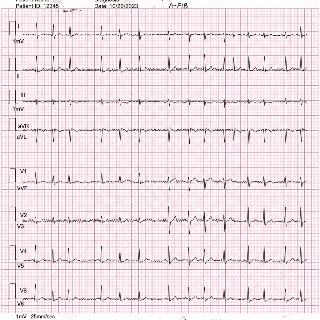
Atrial Fibrillation Rhythm Strip
Classic Atrial Fibrillation electrocardiogram rhythm strip showing an irregularly irregular rhythm, absence of discrete P waves, and variable fibrillatory baseline waves. Rate control is managed first-line with beta-blockers (Metoprolol) or non-dihydropyridine CCBs (Diltiazem); thromboembolic stroke risk is stratified via the CHA2DS2-VASc score, where a score ≥ 2 in men or ≥ 3 in women mandates oral anticoagulation (DOAC preferred over Warfarin).
Heart Failure with Reduced Ejection Fraction (HFrEF) 4-Pillar GDMT Matrix
Clinical Matrix
Pillar ClassRepresentative AgentsMortality Benefit & MechanismMonitoring / Adverse Effects
ARNI / ACEi / ARBSacubitril/Valsartan (preferred), LisinoprilNeprilysin inhibitor + ARB; increases natriuretic peptides, suppresses RAASHyperkalemia, angioedema (36-hr washout required when switching from ACEi to ARNI).
Beta-BlockerCarvedilol, Metoprolol succinate, BisoprololAnti-arrhythmic, reverses cardiac remodeling, blunts chronic sympathetic toxicityBradycardia, heart block; initiate ONLY when euvolemic (never during acute decompensation).
Mineralocorticoid Receptor Antag (MRA)Spironolactone, EplerenoneAldosterone antagonism; prevents myocardial collagen deposition and fibrosisHyperkalemia (K+ > 5.0 mEq/L hold), gynecomastia (Spironolactone; switch to Eplerenone).
SGLT2 InhibitorEmpagliflozin, DapagliflozinPromotes osmotic diuresis, improves myocardial energetics, reduces hospitalizationsEuglycemic DKA, mycotic genital infections, transient eGFR dip (reversible).
Cardiac Autonomics & Chapman Reflex Coordinates
COMLEX & OPP Board Integration
Cardiac sympathetic outflow originates from T1–T5 (T1–T2 SA node, T3–T5 AV node and myocardium). Anterior Chapman point for the myocardium is located at the 2nd intercostal space adjacent to the sternum; the posterior point lies between the spinous and transverse processes of T2 and T3. Somatic dysfunction at T2 on the left strongly correlates with ischemic myocardial strain. *Treatment:* Gentle rib raising and suboccipital decompression normalize sympathetic-parasympathetic tone and improve stroke volume.

1.2 Acute Arrhythmias, Heart Blocks & Valvular Heart Disease

Cardiac dysrhythmias divide into narrow-complex tachycardias (QRS < 120 ms) and wide-complex tachycardias (QRS ≥ 120 ms). In any unstable tachyarrhythmia (hypotension, acute altered mental status, ischemic chest pain, pulmonary edema), the immediate treatment is Synchronized Cardioversion (NOT defibrillation, which is reserved for pulseless VT and VF). In stable narrow-complex supraventricular tachycardia (SVT/AVNRT), vagal maneuvers (modified Valsalva maneuver) are first-line, followed by rapid IV Adenosine (6 mg rapid push followed by 12 mg). Atrial Fibrillation requires immediate assessment of hemodynamic stability, rate vs. rhythm control (Beta-blockers like Metoprolol or CCBs like Diltiazem first-line for rate control; target resting HR < 110 bpm), and thromboembolic risk scoring using CHA2DS2-VASc. A score ≥ 2 in men (≥ 3 in women) mandates therapeutic oral anticoagulation with Direct Oral Anticoagulants (DOACs: Apixaban, Rivaroxaban) preferred over Warfarin.

Bradyarrhythmias and conduction blocks: First-degree AV block (prolonged PR > 200 ms, every P conducts) is benign and requires observation. Second-degree AV block Mobitz Type I (Wenckebach: progressive PR prolongation until a QRS drop) is nodal and usually asymptomatic. Second-degree AV block Mobitz Type II (intermittent dropped QRS without prior PR prolongation; infranodal His-Purkinje failure) carries a high risk of progression to Complete (Third-Degree) Heart Block (complete AV dissociation where P waves and QRS complexes beat independently at intrinsic escape rates 30–40 bpm). Mobitz Type II and Complete Heart Block are class I indications for urgent temporary transcutaneous/transvenous pacing followed by permanent pacemaker implantation.

Valvular Heart Disease: Aortic Stenosis (AS), driven by age-related calcification or congenital bicuspid aortic valve, presents classically with the triad of 'SAD': Syncope, Angina, and Dyspnea on exertion. Physical exam demonstrates a harsh crescendo-decrescendo systolic ejection murmur radiating to the carotids, single or paradoxically split S2, and pulsus parvus et tardus (weak, delayed carotid upstrokes). Symptomatic severe AS (aortic valve area ≤ 1.0 cm², mean gradient ≥ 40 mmHg) carries a 50% 2-year mortality if untreated; definitive management is emergent Transcatheter Aortic Valve Replacement (TAVR) or Surgical Aortic Valve Replacement (SAVR). Mitral Regurgitation presents with a blowing holosystolic murmur at the apex radiating to the axilla; acute severe MR (papillary muscle rupture 2–7 days post-MI) produces sudden cardiogenic pulmonary edema and hypotension requiring emergent surgical repair.

Board Trap — Synchronized Cardioversion vs. Defibrillation
High-Yield Board Trap & Alert
A 58-year-old male with a history of CAD presents with sustained ventricular tachycardia (wide QRS > 120 ms, regular rate 175 bpm). His blood pressure is 78/46 mmHg and he is pale and diaphoresis. An unsynchronized shock (defibrillation) must NOT be delivered! Unsynchronized defibrillation can fire during the vulnerable relative refractory period of the cardiac cycle (the peak or downslope of the T wave), precipitating 'R-on-T' phenomenon and inducing fatal ventricular fibrillation. In any tachyarrhythmia with a palpable pulse (even if hypotensive), you must ALWAYS engage the 'SYNC' button to deliver synchronized electrical cardioversion timed precisely to the R wave.

Chapter 1 Quick-Check Self-Assessment

Test your clinical reasoning before moving to the next chapter
3 Vignettes
Board Vignette #1 Single Best Answer
A 64-year-old male with an acute inferior wall STEMI (leads II, III, aVF) has blood pressure 82/50 mmHg, pulse 108 bpm, and clear lung fields bilaterally. Neck veins are distended to the angle of the jaw. Which of the following is strictly contraindicated in this patient?
Board Vignette #2 Single Best Answer
A 71-year-old female with HFrEF (LVEF 28%) is hospitalized for dyspnea and lower extremity edema. She is stabilized on IV furosemide and her euvolemic baseline is reached. She is currently on Lisinopril 20 mg daily and Spironolactone 25 mg daily. Which medication class must be initiated prior to discharge to reduce long-term all-cause mortality?
Board Vignette #3 Single Best Answer
A 58-year-old male with NYHA Class III HFrEF (LVEF 28%) is currently taking lisinopril 20 mg daily, carvedilol 25 mg twice daily, and spironolactone 25 mg daily. Blood pressure is 118/74 mmHg and HR is 68 bpm. Potassium is 4.4 mEq/L and eGFR is 62 mL/min. What addition to his regimen has been shown to reduce cardiovascular death and heart failure hospitalizations?
Estimated study time: 14 min
Chapter 2 • Gastroenterology & Hepatology
13 min TOC

Gastroenterology, Hepatology & Cirrhosis Decompensation

Ascites & SBP, Hepatic Encephalopathy, Hepatorenal Syndrome, Acute Pancreatitis, and IBD Differentials

Clinical Overview & Board Focus

Gastrointestinal and hepatic pathology spans inflammatory, obstructive, and vascular diseases. Mastery requires calculating and interpreting the Serum-Ascites Albumin Gradient (SAAG), executing spontaneous bacterial peritonitis prophylaxis, applying the Atlanta criteria for acute pancreatitis, and differentiating Crohn's disease from Ulcerative Colitis.

2.1 Cirrhosis Complications: Ascites, SBP & Hepatic Encephalopathy

Cirrhosis results from chronic hepatic injury (alcohol, metabolic dysfunction-associated steatohepatitis [MASH], hepatitis C or B) leading to diffuse parenchymal nodularity and bridging fibrosis. Portal hypertension drives splanchnic vasodilation mediated by excess nitric oxide, producing decreased effective circulating arterial volume, activation of the renin-angiotensin-aldosterone system (RAAS), and renal sodium retention leading to ascites. Diagnostic paracentesis is mandatory for all patients hospitalized with new-onset or worsening ascites. Analysis includes cell count with differential, total protein, and calculation of the Serum-Ascites Albumin Gradient (SAAG = serum albumin minus ascitic fluid albumin). A SAAG ≥ 1.1 g/dL indicates Portal Hypertension (cirrhosis, congestive heart failure, Budd-Chiari syndrome); a SAAG < 1.1 g/dL indicates non-portal causes (peritoneal carcinomatosis, tuberculous peritonitis, nephrotic syndrome, pancreatitis).

Spontaneous Bacterial Peritonitis (SBP) is an acute bacterial infection of ascitic fluid occurring without an identifiable surgically treatable intra-abdominal source, resulting from bacterial translocation across the gut wall. Clinical presentation is often subtle: low-grade fever, mild abdominal pain, encephalopathy, or sudden worsening of renal function. SBP is definitively diagnosed when ascitic fluid absolute neutrophil count is ≥ 250 polymorphonuclear cells (PMNs)/µL. First-line empiric therapy is IV Cefotaxime or Ceftriaxone (2 g IV daily × 5 days). Intravenous Albumin (1.5 g/kg on day 1, and 1.0 g/kg on day 3) is MANDATORY in patients with BUN > 30 mg/dL, creatinine > 1.0 mg/dL, or total bilirubin > 4 mg/dL to prevent acute renal failure from type 1 hepatorenal syndrome and reduce mortality.

Hepatic Encephalopathy (HE) is a neurocognitive syndrome caused by failure of the cirrhotic liver to detoxify gut-derived neurotoxins, predominantly ammonia. It manifests along the West Haven criteria from subtle sleep-wake cycle reversal to asterixis ('flapping tremor' on dorsiflexion of wrists), confusion, and coma. HE is frequently precipitated by an acute stressor: GI bleeding (blood acts as a protein load), constipation, infection/SBP, electrolyte disturbances (hypokalemia promotes renal ammoniagenesis), or sedatives. First-line therapy is Lactulose (titrated to achieve 2–3 soft bowel movements daily; converted by colonic bacteria into lactic acid, trapping NH4+ in stool); Rifaximin is added as second-line therapy for recurrent episodes.

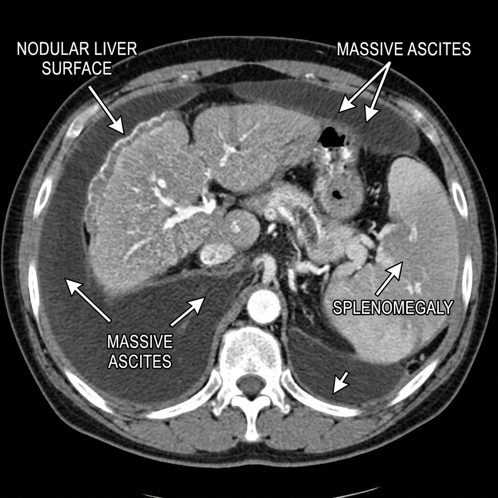
Cirrhosis with Ascites and Splenomegaly CT
Contrast-enhanced abdominal CT demonstrating severe decompensated cirrhosis: Note the shrunken, nodular liver contour, massive low-attenuation ascites fluid surrounding the liver and spleen, and prominent splenomegaly secondary to portal hypertension. Diagnostic paracentesis is mandatory to calculate the serum-ascites albumin gradient (SAAG ≥ 1.1 g/dL indicates portal hypertension) and rule out spontaneous bacterial peritonitis (PMNs ≥ 250/µL).
Inflammatory Bowel Disease Comparison Matrix
Clinical Matrix
FeatureCrohn's DiseaseUlcerative Colitis (UC)
Anatomic DistributionAny segment from mouth to anus; skip lesions; terminal ileum #1Continuous involvement starting at rectum, extending proximally; colon only
Depth of InflammationTransmural (mucosa through serosa)Mucosal and submucosal only
Endoscopic AppearanceCobblestoning, aphthous and deep longitudinal ulcers, creeping fatContinuous erythema, friability, loss of vascular markings, pseudopolyps
Histologic HallmarkNon-caseating granulomas (pathognomonic in 30%)Crypt abscesses with neutrophils, architectural distortion
ComplicationsFistulas (enterovesical, enterocutaneous), strictures, abscesses, perianal diseaseToxic Megacolon (> 6 cm colonic dilation), high risk of colorectal cancer
Serologic MarkersPositive ASCA (anti-Saccharomyces cerevisiae antibody)Positive p-ANCA (perinuclear antineutrophil cytoplasmic antibody)
Smoking RelationshipSmoking INCREASES risk and worsens disease courseSmoking is PROTECTIVE; cessation frequently precipitates flare
Board Trap — Toxic Megacolon Contraindications
High-Yield Board Trap & Alert
A 26-year-old with a severe flare of Ulcerative Colitis develops high fever (39.2°C), marked tachycardia (128 bpm), hypotension, abdominal distention, and leukocytosis (24,000/µL). Abdominal radiograph demonstrates transverse colon dilation to 7.5 cm. Complete colonoscopy and antimotility agents (loperamide, narcotics) are STRICTLY CONTRAINDICATED due to the extreme risk of colonic perforation and peritonitis. Management requires NPO, NG tube decompression, IV fluids, IV Methylprednisolone, broad-spectrum IV antibiotics, and urgent surgical consult for subtotal colectomy if not improving within 24–48 hours.

2.2 Acute Pancreatitis & Peptic Ulcer Disease

Acute Pancreatitis diagnosis requires ≥ 2 of the 3 revised Atlanta criteria: (1) Severe, persistent epigastric pain radiating directly to the mid-back; (2) Serum lipase or amylase ≥ 3× upper limit of normal (lipase is superior: more sensitive, specific, and remains elevated for 8–14 days); and (3) Characteristic findings on contrast-enhanced abdominal CT or MRI. If classic pain and ≥ 3× lipase elevation are present, diagnostic CT is NOT required on admission and should be deferred unless diagnostic uncertainty or failure to improve after 48–72 hours raises concern for pancreatic necrosis, pseudocyst, or abscess.

Initial management focuses on aggressive isotonic intravenous fluid resuscitation (Lactated Ringer's solution preferred over normal saline; 200–500 mL/hr, or 5–10 mL/kg/hr) to maintain adequate microvascular perfusion of the pancreatic parenchyma and prevent necrosis, monitoring urine output (> 0.5 mL/kg/hr) and hematocrit reduction. Mild acute pancreatitis warrants early oral feeding (low-fat solid or liquid diet as tolerated) as soon as abdominal pain improves and ileus resolves. Routine prophylactic intravenous antibiotics are NOT recommended for acute or sterile necrotizing pancreatitis; antibiotics (Meropenem or Ciprofloxacin + Metronidazole) are reserved strictly for confirmed infected necrosis (CT showing retroperitoneal gas bubbles or positive fine-needle aspirate gram stain/culture).

Peptic Ulcer Disease (PUD) is driven predominantly by Helicobacter pylori infection (> 70% duodenal, 50% gastric) and non-steroidal anti-inflammatory drugs (NSAIDs, which inhibit COX-1-mediated mucosal prostaglandin E2 synthesis). Duodenal ulcers classically cause epigastric pain relieved by food or antacids, recurring 2–4 hours after meals and at night. Gastric ulcers present with pain exacerbated by food intake, leading to food avoidance and weight loss. All gastric ulcers require follow-up endoscopy with repeat biopsy in 6–8 weeks to rule out underlying gastric adenocarcinoma. First-line eradication for H. pylori is Bismuth Quadruple Therapy × 14 days: Proton Pump Inhibitor (Omeprazole BID) + Bismuth subsalicylate + Metronidazole + Tetracycline. Eradication MUST be verified ≥ 4 weeks post-treatment using a urea breath test or stool antigen assay (patient must be off PPI for ≥ 2 weeks prior to testing).

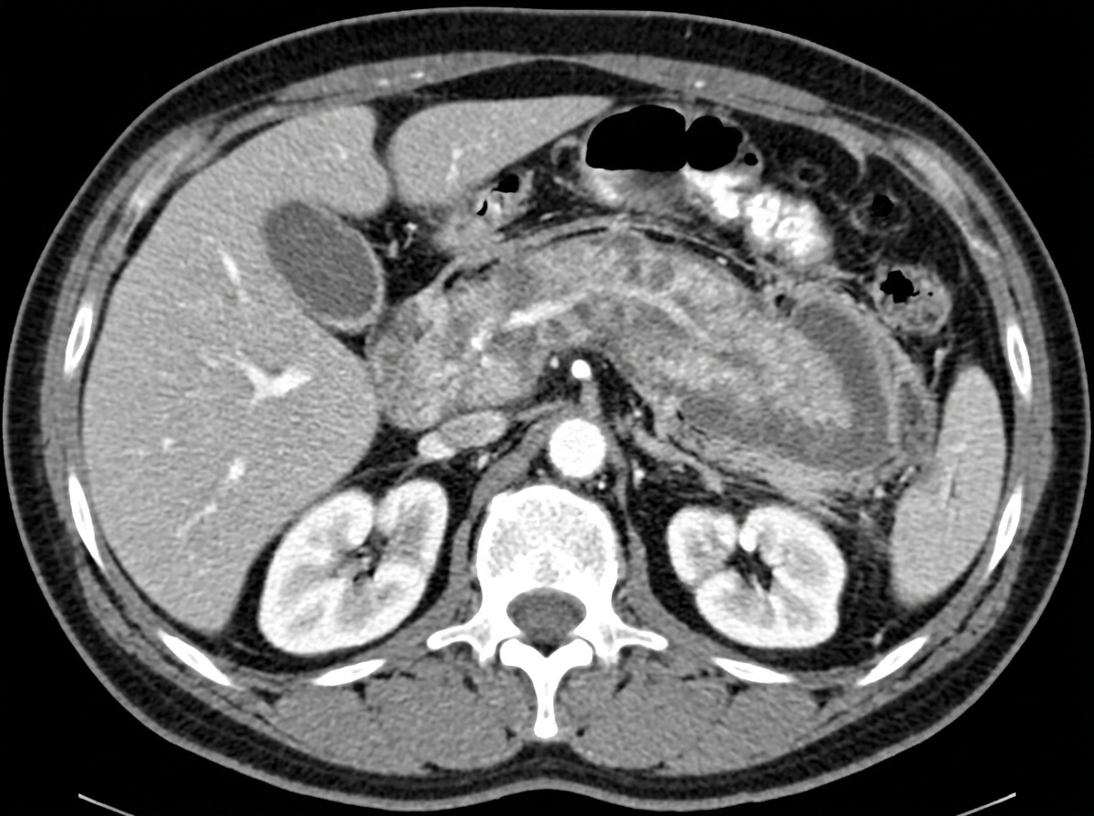
Acute Pancreatitis Abdominal CT
Contrast-enhanced axial abdominal CT demonstrating acute pancreatitis: Diffuse enlargement and edema of the pancreatic body and tail with extensive peripancreatic fat stranding and fluid tracking into the anterior pararenal space. Diagnosis requires ≥ 2 of 3 revised Atlanta criteria: epigastric pain radiating to back, lipase/amylase ≥ 3× ULN, and characteristic imaging findings.
COMLEX Clinical Integration — Splanchnic Collateral Reflexes in Foregut Disease
COMLEX & OPP Board Integration
The stomach, duodenum, liver, and pancreas share sympathetic preganglionic innervation from T5 to T9 via the greater splanchnic nerve, synapsing in the celiac ganglion. Viscerosomatic facilitation produces bilateral paraspinal hypertonicity, boggy tissue changes, and tenderness at T5–T9 (frequently left-sided for stomach/pancreas, right-sided for liver/duodenum). Anterior Chapman's point for the stomach is located in the 5th and 6th left intercostal spaces midclavicular line; the pancreatic Chapman's point is at the 7th right intercostal space. Collateral ganglion release (celiac ganglion release anteriorly between xiphoid and umbilicus) and gentle paraspinal inhibition relieve splanchnic vasoconstriction, promote mucosal perfusion, and accelerate ulcer healing.

Chapter 2 Quick-Check Self-Assessment

Test your clinical reasoning before moving to the next chapter
3 Vignettes
Board Vignette #1 Single Best Answer
A 52-year-old male with alcoholic cirrhosis is hospitalized with low-grade fever (38.1°C) and abdominal distention. Paracentesis reveals straw-colored ascitic fluid with total protein 1.1 g/dL, serum albumin 3.0 g/dL, ascitic albumin 0.8 g/dL, and neutrophil count 340 PMNs/µL. What is the mandatory next medical step?
Board Vignette #2 Single Best Answer
A 45-year-old female presents with severe constant epigastric pain radiating to her back, accompanied by vomiting. Serum lipase is 1,450 U/L (normal < 60 U/L). Abdominal exam reveals epigastric tenderness without rebound or guarding. What is the most appropriate next step in management?
Board Vignette #3 Single Best Answer
A 66-year-old male with a 45-pack-year smoking history presents with worsening dyspnea and productive cough. Spirometry demonstrates FEV1/FVC 0.58 and post-bronchodilator FEV1 42% of predicted. He has suffered 2 COPD exacerbations requiring oral corticosteroids in the past year. What is the most appropriate long-term maintenance inhaled regimen?
Estimated study time: 13 min
Chapter 3 • Nephrology & Acid-Base
15 min TOC

Nephrology, Acute Kidney Injury & Electrolyte Disorders

Prerenal vs. Intrinsic AKI, Glomerulonephritis, Hyponatremia (Osmotic Demyelination), and Hyperkalemia Protocols

Clinical Overview & Board Focus

Renal and electrolyte disorders require systematic algorithmic approaches. Mastery encompasses differentiating prerenal azotemia from acute tubular necrosis (ATN), interpreting urine microscopy (casts), managing glomerulonephritis, correcting sodium derangements without causing brain damage, and deploying multi-step hyperkalemia stabilization.

3.1 Acute Kidney Injury (AKI) & Electrolyte Homeostasis

Acute Kidney Injury (AKI) is classified into Prerenal, Intrinsic, and Postrenal etiologies. Prerenal azotemia results from hypoperfusion (hypovolemia, sepsis, heart failure, NSAIDs constricting afferent arterioles, ACE inhibitors dilating efferent arterioles). The renal tubules remain intact, avidly reabsorbing sodium and water: BUN/Creatinine ratio > 20:1, Urine Sodium < 20 mEq/L, Fractional Excretion of Sodium (FeNa) < 1% (or FeUrea < 35% if on loop diuretics), and urine specific gravity > 1.020 with hyaline casts. Intrinsic AKI is predominantly Acute Tubular Necrosis (ATN) caused by prolonged renal ischemia or nephrotoxins (aminoglycosides, IV iodinated contrast, vancomycin, cisplatin, myoglobin from rhabdomyolysis). In ATN, tubular epithelium is damaged, losing concentrated reabsorption: BUN/Cr ratio 10–15:1, Urine Sodium > 40 mEq/L, FeNa > 2%, and pathognomonic muddy brown granular casts.

Hyponatremia (serum sodium < 135 mEq/L) evaluation begins with serum osmolality to exclude Hypertonic (hyperglycemia; add 1.6–2.0 mEq/L Na for every 100 mg/dL glucose > 100) and Pseudohyponatremia (severe hyperlipidemia or paraproteinemia). True hypotonic hyponatremia is categorized by volume status: Hypovolemic (dehydration, diuretics; urine Na < 20 if extrarenal, > 20 if renal), Hypervolemic (CHF, cirrhosis, nephrotic syndrome), and Euvolemic (SIADH, primary polydipsia, secondary adrenal insufficiency, hypothyroidism). In SIADH, urine osmolality is inappropriately concentrated (> 100 mOsm/kg, usually > 300) and urine sodium is high (> 40 mEq/L). Safe correction is Paramount: serum sodium must NOT be corrected faster than 6 to 8 mEq/L in 24 hours to prevent Central Pontine Myelinolysis (Osmotic Demyelination Syndrome), which presents days later with dysarthria, dysphagia, quadriparesis, and 'locked-in' syndrome.

Hyperkalemia (serum potassium > 5.0 mEq/L; severe ≥ 6.5 mEq/L) represents an immediate cardiac electrical emergency. Progressive ECG changes evolve from tall, peaked T waves (earliest finding) to PR prolongation, loss of P waves, widening of the QRS complex, 'sine-wave' pattern, and ventricular fibrillation or asystole. Emergency management follows three sequential steps: (1) Cardiac membrane stabilization: IV Calcium Gluconate (10 mL of 10% solution over 2–3 minutes; stabilizes the cardiac sarcolemma within minutes without lowering serum potassium); (2) Intracellular potassium shifting: IV regular Insulin (10 units) with 50% Dextrose (50 mL D50), nebulized Albuterol (10–20 mg), and IV Sodium Bicarbonate (if metabolic acidosis present); and (3) Total body potassium elimination: Loop diuretics (Furosemide), potassium binders (Patiromer or Sodium Zirconium Cyclosilicate [Lokelma]), and emergent Hemodialysis for refractory hyperkalemia.

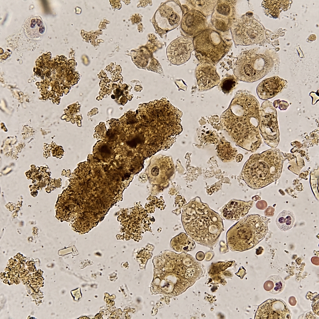
Muddy Brown Granular Casts Acute Tubular Necrosis
High-power urine microscopy demonstrating classic muddy brown granular casts composed of degenerated renal tubular epithelial cells embedded in Tamm-Horsfall protein matrix. This finding is pathognomonic for Acute Tubular Necrosis (ATN), characterized by impaired tubular sodium reabsorption (FeNa > 2%, Urine Na > 40 mEq/L, BUN/Cr ratio < 15:1).
Electrolyte Correction Velocity & Toxic Syndromes
Clinical Matrix
Electrolyte EmergencyAcute Clinical ManifestationKey InterventionLethal Iatrogenic Pitfall
Severe Hyponatremia (< 120 mEq/L)Cerebral edema, seizures, coma3% Hypertonic Saline (100 mL boluses) for seizuresRapid correction > 8 mEq/L/day causes Osmotic Demyelination Syndrome (locked-in)
Severe Hypernatremia (> 150 mEq/L)Lethargy, irritability, intracranial hemorrhageD5W or 0.45% NS calculated via Free Water DeficitRapid correction > 10 mEq/L/day causes Fatal Cerebral Edema
Severe Hyperkalemia (≥ 6.5 mEq/L)Sine-wave ECG, flaccid paralysis, VFIV Calcium Gluconate → Insulin + D50 → Lokelma / DialysisGiving insulin without calcium gluconate when QRS is widened leads to arrest
Severe Hypokalemia (< 2.5 mEq/L)U waves, flattened T waves, PACs/PVCs, ileusOral/IV Potassium Chloride + IV Magnesium repletionFailure to correct hypomagnesemia causes refractory hypokalemia (ROMK uninhibited)
COMLEX Clinical Integration — Renal Autonomic Innervation & Collateral Release
COMLEX & OPP Board Integration
The kidneys receive sympathetic preganglionic innervation from spinal segments T10 to T11 via the lesser splanchnic nerve, synapsing in the superior mesenteric and aorticorenal ganglia. Sustained sympathetic hyperactivity causes afferent arteriolar vasoconstriction, diminishing glomerular filtration rate and activating renin release. Paraspinal examination reveals acute tissue texture changes and hypertonicity at T10–T11. Chapman's reflex point for the kidney is located 1 inch lateral and 1 inch superior to the umbilicus anteriorly, and between the transverse processes of T12 and L1 posteriorly. Inhibitory pressure at T10–T11 and gentle release of the thoracolumbar junction reduce renal sympathetic tone, promote medullary blood flow, and enhance diuresis.

3.2 Glomerulonephritis, Nephrotic Syndrome & Acid-Base Balance

Glomerular disease partitions into Nephrotic syndromes (massive proteinuria > 3.5 g/day, hypoalbuminemia < 3.0 g/dL, generalized edema, hyperlipidemia, and lipiduria with 'Maltese cross' oval fat bodies) and Nephritic syndromes (hematuria with dysmorphic RBCs and RBC casts, variable sub-nephrotic proteinuria, hypertension, and azotemia). In adults, the most common nephrotic syndromes are Focal Segmental Glomerulosclerosis (FSGS: associated with African-American race, HIV, heroin use, obesity; podocyte effacement on EM), Membranous Nephropathy (associated with anti-PLA2R antibodies, hepatitis B/C, NSAIDs, solid tumors; 'spike-and-dome' subepithelial IgG/C3 deposits), and Diabetic Nephropathy (mesangial expansion and Kimmelstiel-Wilson nodular glomerulosclerosis).

Nephritic syndromes in adults: IgA Nephropathy (Berger disease) is the most common glomerulonephritis worldwide, presenting with recurrent macroscopic hematuria 1–2 days following a viral upper respiratory or GI infection ('synpharyngitic', distinguished from post-streptococcal GN which occurs 1–3 weeks after pharyngitis); renal biopsy demonstrates mesangial IgA and C3 deposition. Rapidly Progressive Glomerulonephritis (RPGN) is characterized by rapid loss of renal function over days to weeks with histologic cellular crescents (macrophages and fibrin) in Bowman's space. RPGN is categorized by immunofluorescence: (1) Linear anti-GBM (Goodpasture syndrome: anti-type IV collagen antibodies, pulmonary alveolar hemorrhage with hemoptysis and renal failure); (2) Granular immune-complex (SLE class IV lupus nephritis, PSGN); and (3) Pauci-immune negative IF (systemic ANCA vasculitides: Granulomatosis with Polyangiitis [c-ANCA / PR3], Microscopic Polyangiitis [p-ANCA / MPO]).

Acid-Base Interpretation follows a three-step rule: (1) Evaluate pH (acidemia < 7.35, alkalemia > 7.45); (2) Identify primary disorder by examining PaCO2 and HCO3-; and (3) Calculate the Anion Gap (AG = Na - [Cl + HCO3]; normal 8–12 mEq/L). In High Anion Gap Metabolic Acidosis (HAGMA), remember the mnemonic MUDPILES: Methanol, Uremia, DKA, Propylene glycol, Isoniazid/Iron, Lactic acidosis, Ethylene glycol, Salicylates. Check respiratory compensation in metabolic acidosis using Winter's formula: expected PaCO2 = 1.5 × HCO3- + 8 ± 2. If measured PaCO2 is higher than expected, a concomitant primary respiratory acidosis is present; if lower, a concomitant primary respiratory alkalosis is present.

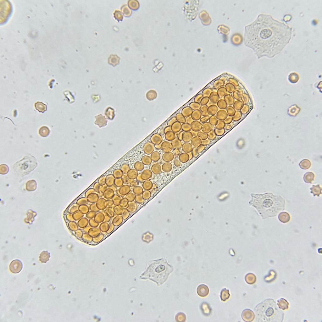
Red Blood Cell Cast Glomerulonephritis
High-power urine microscopy demonstrating a Red Blood Cell (RBC) Cast with dysmorphic erythrocytes trapped in a cylindrical protein matrix. RBC casts are pathognomonic for active Glomerulonephritis (e.g., IgA nephropathy, lupus nephritis, post-infectious GN, or anti-GBM disease), signifying disruption of the glomerular capillary wall and basement membrane.
Board Trap — Berger Disease vs. Post-Streptococcal GN Timeline
High-Yield Board Trap & Alert
A 22-year-old college student develops gross hematuria and sore throat on the SAME day (synpharyngitic hematuria). This is IgA Nephropathy (Berger disease), NOT post-streptococcal glomerulonephritis! Post-Streptococcal Glomerulonephritis (PSGN) requires a latency period of 1 to 3 weeks following streptococcal pharyngitis (or 3 to 6 weeks following impetigo) for immune complex formation and glomerular deposition to occur. In PSGN, serum complement C3 is low; in IgA nephropathy, complement C3 is completely normal.

Chapter 3 Quick-Check Self-Assessment

Test your clinical reasoning before moving to the next chapter
3 Vignettes
Board Vignette #1 Single Best Answer
A 68-year-old male with chronic hyponatremia is hospitalized with sodium of 114 mEq/L. Over the first 24 hours of vigorous 3% saline therapy, his repeat serum sodium is 132 mEq/L (an increase of 18 mEq/L). Three days later, he becomes mute, quadriplegic, and cannot swallow, but can move his eyes vertically. What iatrogenic complication has occurred?
Board Vignette #2 Single Best Answer
A 24-year-old male presents with gross hematuria, hemoptysis, and acute renal failure with creatinine 4.2 mg/dL. Renal biopsy reveals cellular crescents in Bowman's space and linear IgG staining along the glomerular basement membrane. What is the definitive diagnosis and primary therapy?
Board Vignette #3 Single Best Answer
A 52-year-old male with decompensated alcoholic cirrhosis presents with fever (101.4°F), abdominal distension, and diffuse tenderness without peritoneal signs. Diagnostic paracentesis reveals cloudy ascitic fluid with neutrophil count of 420 cells/mm3 and total protein of 1.1 g/dL. What is the definitive initial management?
Estimated study time: 15 min
Chapter 4 • Endocrinology & Metabolism
13 min TOC

Endocrinology, Diabetes Emergencies & Endocrine Crises

DKA vs. HHS Protocols, Thyroid Storm & Myxedema Coma, Adrenal Crisis, and Calcium Disorders

Clinical Overview & Board Focus

Endocrine emergencies feature extreme metabolic derangements requiring multi-phase fluid, electrolyte, and hormone replacement algorithms. Board examinations demand fluency in the distinction between DKA and HHS, management of thyroid storm, and localization of Cushing and Addison syndromes.

4.1 Diabetic Ketoacidosis (DKA), HHS & Endocrine Crises

Diabetic Ketoacidosis (DKA) is a life-threatening complication predominantly occurring in Type 1 Diabetes Mellitus, triggered by absolute insulin deficiency coupled with counter-regulatory hormone excess (glucagon, epinephrine, cortisol). Diagnostic triad: Hyperglycemia (blood glucose > 250 mg/dL), Anion Gap Metabolic Acidosis (pH < 7.30, serum bicarbonate < 18 mEq/L, anion gap > 12), and Ketonemia/Ketonuria (elevated beta-hydroxybutyrate). Patients present with Kussmaul respirations (rapid deep breathing to blow off CO2), fruity acetone breath, vomiting, and abdominal pain. Hyperosmolar Hyperglycemic State (HHS) occurs in Type 2 Diabetes, characterized by severe hyperglycemia (> 600 mg/dL), high serum osmolality (> 320 mOsm/kg), and profound dehydration (fluid deficit 8–10 L) in the ABSENCE of significant ketoacidosis (pH > 7.30, bicarb > 18).

DKA Resuscitation Algorithm: (1) Fluid resuscitation: 0.9% Normal Saline at 1 L/hr for the first 1–2 hours. Once hemodynamically stable, switch to 0.45% NS if corrected sodium is normal or high; (2) Potassium protocol: Insulin drives potassium into cells. If serum K < 3.3 mEq/L, HOLD insulin and replete potassium (20–40 mEq/hr) until K ≥ 3.3 mEq/L to prevent fatal cardiac arrest; if K is 3.3–5.2 mEq/L, give insulin AND add 20–30 mEq K per liter of IV fluid; if K > 5.2 mEq/L, give insulin and check K every 2 hours; (3) Regular Insulin infusion: 0.1 units/kg IV bolus followed by 0.1 units/kg/hr infusion (or 0.14 units/kg/hr continuous). When blood glucose drops to 200 mg/dL in DKA (or 300 mg/dL in HHS), add 5% Dextrose (D5 with 0.45% NS) to the infusion while continuing insulin to close the anion gap without precipitating hypoglycemia. DKA is resolved when: glucose < 200 mg/dL PLUS two of: serum bicarb ≥ 15 mEq/L, venous pH > 7.30, and anion gap ≤ 12.

Thyroid Storm is a hypermetabolic crisis in uncontrolled hyperthyroidism (Graves disease, toxic multinodular goiter), triggered by infection, trauma, or iodinated contrast. Patients present with high fever (> 38.5°C), marked tachycardia (> 140 bpm, atrial fibrillation), high-output heart failure, delirium, and diarrhea. Management requires four sequential pharmacologic steps: (1) Beta-blocker: Propranolol (blocks adrenergic hyperactivity and peripheral T4-to-T3 conversion); (2) Thionamide: Propylthiouracil (PTU, preferred in thyroid storm as it blocks new thyroid hormone synthesis AND peripheral T4-to-T3 conversion; Methimazole is second-line); (3) Saturated Solution of Potassium Iodide (SSKI / Lugol solution): administered at least 1 hour AFTER PTU to block hormone release via the Wolff-Chaikoff effect (giving iodine first worsens thyroid storm by providing substrate!); and (4) IV Glucocorticoids: Hydrocortisone (blocks peripheral T4-to-T3 conversion and treats relative adrenal insufficiency).

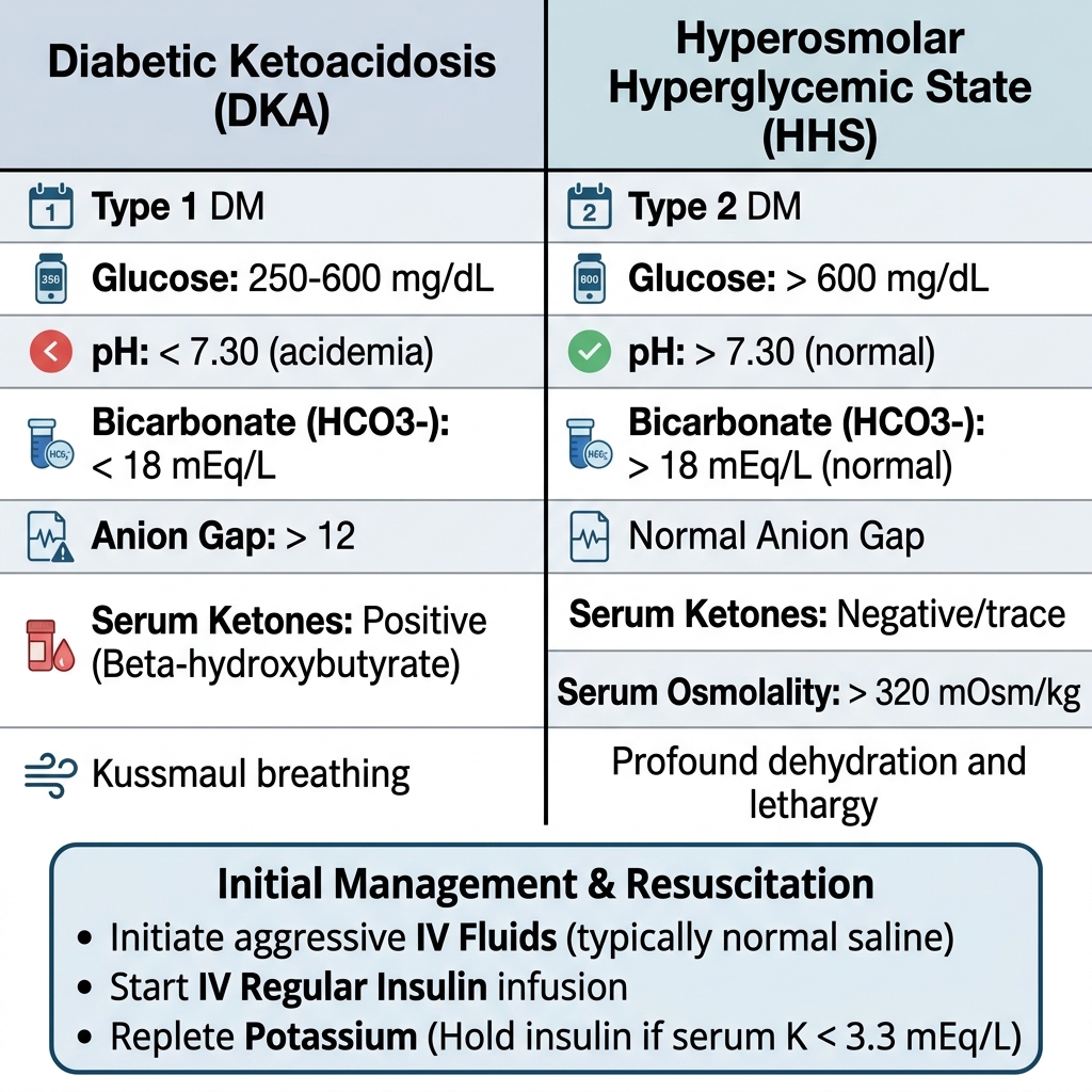
DKA vs HHS Diagnostic and Management Comparison
Diagnostic and resuscitation comparison matrix between Diabetic Ketoacidosis (DKA) and Hyperosmolar Hyperglycemic State (HHS). In DKA, insulinopenia triggers uninhibited lipolysis and ketogenesis, causing high anion gap metabolic acidosis. In HHS, residual insulin prevents ketoacidosis but allows severe hyperglycemia (>600 mg/dL) and hyperosmolality (>320 mOsm/kg). Initial priority for both: aggressive IV isotonic crystalloids and checking potassium before insulin.
Acute Kidney Injury (AKI) Laboratory Differentiation Matrix
Clinical Matrix
Diagnostic ParameterPrerenal AzotemiaIntrinsic Renal (ATN)Postrenal (Obstruction)
BUN : Creatinine Ratio> 20 : 1 (urea reabsorption avid)< 15 : 1 (tubular backleak)Variable (initially > 15:1)
Fractional Excretion of Na (FeNa)< 1% (intact sodium retention)> 2% (tubular epithelial failure)> 2% (late obstruction)
Urine Sodium Concentration< 20 mEq/L> 40 mEq/L> 40 mEq/L (late)
Urine Osmolality> 500 mOsm/kg (concentrated)< 350 mOsm/kg (isosthenuria)< 350 mOsm/kg
Urine Sediment MicroscopyHyaline casts, bland sedimentMuddy brown granular casts, renal tubular cellsNormal or RBCs/WBCs/crystals
Board Trap — Order of Treatment in Thyroid Storm
High-Yield Board Trap & Alert
Always administer the thionamide (PTU or Methimazole) at least 60 minutes BEFORE administering Lugol's iodine or potassium iodide in thyroid storm! If inorganic iodine is administered prior to blocking organification with PTU, the thyroid gland will rapidly incorporate the exogenous iodine into thyroglobulin, synthesizing massive new stores of T4 and T3 (Jod-Basedow phenomenon), acutely worsening the thyroid crisis.

Chapter 4 Quick-Check Self-Assessment

Test your clinical reasoning before moving to the next chapter
3 Vignettes
Board Vignette #1 Single Best Answer
A 23-year-old female with Type 1 Diabetes presents to the ED with severe vomiting and confusion. Vitals: BP 92/58 mmHg, HR 122 bpm, RR 32/min (Kussmaul). Point-of-care fingerstick glucose is 480 mg/dL. Initial BMP reveals Na 130 mEq/L, K 2.9 mEq/L, Cl 96 mEq/L, HCO3 10 mEq/L, venous pH 7.15. What is the most critical first pharmacological step?
Board Vignette #2 Single Best Answer
A 36-year-old female with Graves disease presents with high fever (40.1°C), atrial fibrillation with ventricular response of 165 bpm, extreme agitation, and jaundice following an urgent dental extraction. After starting IV propranolol, in what sequence should the next pharmacologic agents be given?
Board Vignette #3 Single Best Answer
A 48-year-old female presents with hemoptysis, dyspnea, and oliguria. Serum creatinine is 4.6 mg/dL. Urinalysis shows 3+ protein, dysmorphic RBCs, and RBC casts. Renal biopsy demonstrates cellular crescents in 70% of glomeruli and linear IgG staining on direct immunofluorescence. What is the underlying pathogenesis?
Estimated study time: 13 min
Chapter 5 • Hematology & Oncology
14 min TOC

Hematology, Transfusion Medicine & Oncologic Emergencies

Microcytic vs. Macrocytic Anemias, Coagulation Disorders, TTP vs. ITP, and Tumor Lysis Syndrome

Clinical Overview & Board Focus

Hematologic diagnosis relies heavily on peripheral blood smear morphology, red cell indices (MCV, RDW), and coagulation profiles (PT/INR, PTT). Oncologic emergencies (tumor lysis syndrome, spinal cord compression, hypercalcemia of malignancy) represent critical internal medicine clinical priorities.

5.1 Microcytic Anemia Differential & Thrombotic Thrombocytopenic Purpura

Microcytic Anemia (MCV < 80 fL) is categorized via iron studies: Iron Deficiency Anemia (IDA) is marked by low serum iron, elevated total iron-binding capacity (TIBC / transferrin), low ferritin (< 30 ng/mL; the most sensitive test), and elevated red cell distribution width (RDW). In men and postmenopausal women, IDA is colon cancer until proven otherwise, mandating upper endoscopy and colonoscopy. Anemia of Chronic Disease (ACD) is driven by IL-6 induced hepatic production of hepcidin (which internalizes ferroportin, sequestering iron inside macrophages): low serum iron, low or normal TIBC, normal or elevated ferritin (> 100 ng/mL), and normal RDW. Thalassemia Trait is characterized by profound microcytosis (MCV often < 70 fL) with completely normal iron studies, normal or elevated RBC count, low RDW (Mentzer index: MCV/RBC < 13 suggests thalassemia; > 13 suggests IDA), and abnormal hemoglobin electrophoresis (elevated HbA2 > 3.5% in beta-thalassemia minor). Sideroblastic Anemia (alcohol, lead toxicity, isoniazid/B6 deficiency) shows elevated serum iron and ferritin with ringed sideroblasts on Prussian blue bone marrow staining.

Thrombotic Thrombocytopenic Purpura (TTP) is a hematologic emergency resulting from severe deficiency of ADAMTS13 (a metalloproteinase that cleaves ultra-large von Willebrand factor multimers), usually caused by an acquired IgG autoantibody. Uncleaved vWF multimers promote massive platelet microthrombi in small vessels, causing end-organ ischemia and shearing erythrocytes. The classic pentad is: (1) Microangiopathic Hemolytic Anemia (MAHA: schistocytes on blood smear, high LDH, low haptoglobin, negative Coombs); (2) Severe Thrombocytopenia (often < 20,000/µL); (3) Neurologic deficits (confusion, headache, focal deficits); (4) Renal dysfunction; and (5) Fever. PT, PTT, and fibrinogen are strictly NORMAL (distinguishing TTP from DIC, which has prolonged PT/PTT and low fibrinogen). Immediate treatment is emergent Plasma Exchange (PLEX / plasmapheresis) combined with high-dose systemic Corticosteroids and Caplacizumab (anti-vWF nanobody). Platelet transfusions are STRICTLY CONTRAINDICATED as they fuel widespread microthrombosis.

Tumor Lysis Syndrome (TLS) is an oncologic emergency occurring after initiation of cytotoxic chemotherapy for high-turnover hematologic malignancies (Burkitt lymphoma, ALL, AML). Massive cell lysis floods circulation with intracellular contents, producing the classic metabolic tetrad: Hyperkalemia, Hyperphosphatemia, Hyperuricemia, and secondary Hypocalcemia (precipitation of calcium phosphate crystals in renal tubules). Patients develop acute uric acid nephropathy, cardiac arrhythmias, and tetany. Prophylaxis and treatment require aggressive IV hydration, Rasburicase (recombinant urate oxidase that converts uric acid to soluble allantoin; drug of choice in established TLS or high risk), or Allopurinol (xanthine oxidase inhibitor, used for low/moderate risk prophylaxis).

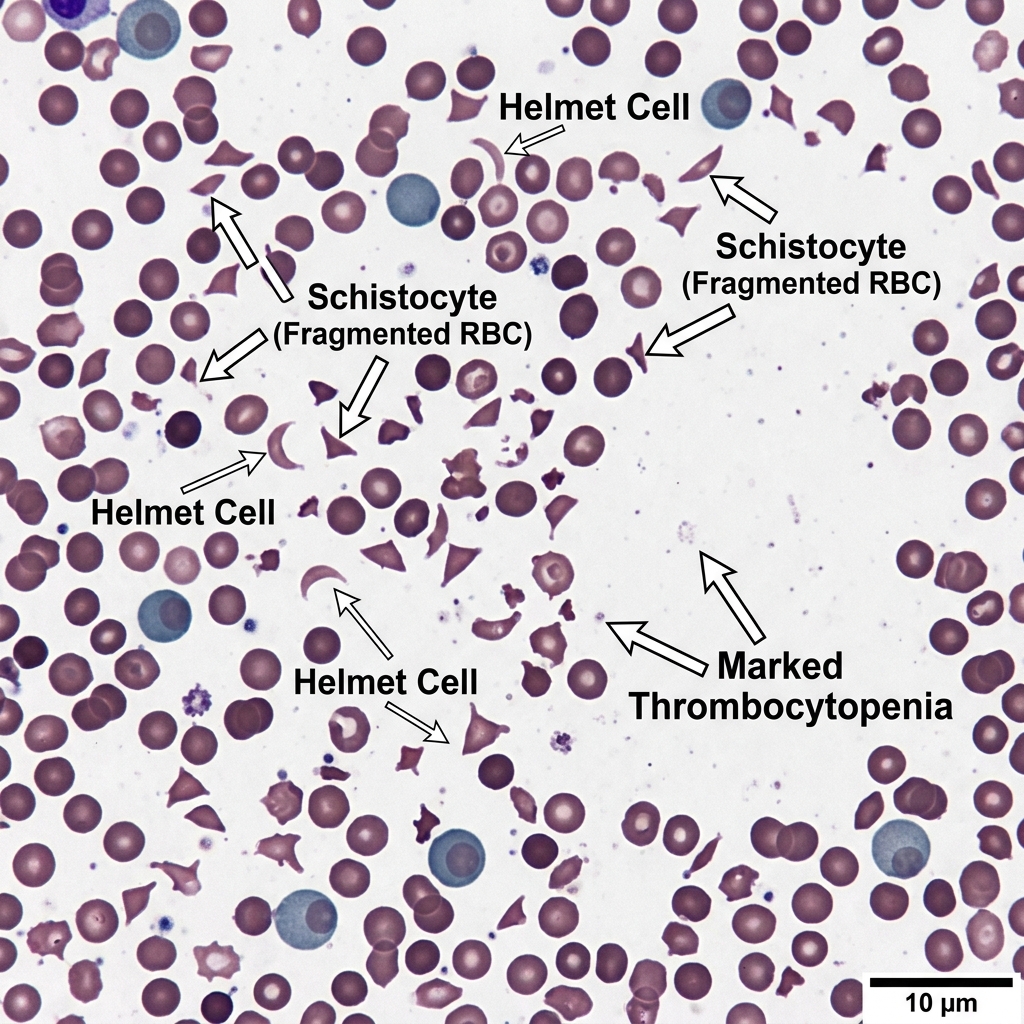
Schistocytes and Helmet Cells Peripheral Blood Smear
High-power peripheral blood smear demonstrating numerous schistocytes (fragmented red cells) and helmet cells alongside marked thrombocytopenia, characteristic of Microangiopathic Hemolytic Anemia (MAHA) in Thrombotic Thrombocytopenic Purpura (TTP) or Hemolytic Uremic Syndrome (HUS). Shearing occurs as RBCs navigate microvascular platelet thrombi formed due to severe ADAMTS13 deficiency. Coagulation profile (PT, PTT, fibrinogen) remains normal.
Coagulation & Thrombocytopenic Disorders Matrix
Clinical Matrix
DiseasePlatelet CountPT / INRaPTTBleeding TimePathognomonic Finding / Treatment
Immune Thrombocytopenia (ITP)Profoundly Low (< 30k)NormalNormalProlongedAnti-GpIIb/IIIa antibodies; IVIG, Corticosteroids, Rituximab, TPO receptor agonists
TTPLow (< 20k)NormalNormalProlongedSchistocytes; ADAMTS13 < 10%; Emergent Plasma Exchange (PLEX); NO platelets!
Disseminated Intravascular Coag (DIC)LowProlonged (↑)Prolonged (↑)ProlongedLow fibrinogen, elevated D-dimer, schistocytes; treat underlying sepsis/trauma + cryo/FFP
Hemophilia A (Factor VIII def)NormalNormalProlonged (↑↑)NormalX-linked recessive; hemarthrosis; mixing study corrects; Factor VIII concentrate
von Willebrand Disease (vWD)Normal (or mild ↓)NormalNormal or ProlongedProlongedAutosomal dominant; mucosal bleeding; ristocetin cofactor assay abnormal; Desmopressin (DDAVP)
Board Trap — Platelet Transfusions in TTP and HIT
High-Yield Board Trap & Alert
A 34-year-old female presents with confusion, fever, platelets of 12,000/µL, and schistocytes on blood smear. The intern immediately orders a unit of platelets. STOP! Transfusing platelets in Thrombotic Thrombocytopenic Purpura (TTP) or Heparin-Induced Thrombocytopenia (HIT) is 'adding fuel to the fire', provoking catastrophic widespread microvascular thrombosis, acute myocardial infarction, stroke, and death. Platelets are only transfused in TTP if life-threatening catastrophic intracranial hemorrhage is actively underway.

Chapter 5 Quick-Check Self-Assessment

Test your clinical reasoning before moving to the next chapter
3 Vignettes
Board Vignette #1 Single Best Answer
A 31-year-old female presents with acute confusion, fever (38.6°C), and petechiae on her lower extremities. Labs demonstrate Hb 7.8 g/dL, platelets 14,000/µL, LDH 1,200 U/L, haptoglobin < 10 mg/dL, and Cr 2.1 mg/dL. Coagulation profile shows PT 12 sec, INR 1.0, PTT 28 sec, and fibrinogen 320 mg/dL. Peripheral blood smear reveals numerous schistocytes. What is the most appropriate next step?
Board Vignette #2 Single Best Answer
A 19-year-old male with newly diagnosed Burkitt lymphoma starts induction chemotherapy. Twelve hours later, labs reveal K 6.8 mEq/L, PO4 7.2 mg/dL, Ca 6.9 mg/dL, Uric acid 14.2 mg/dL, and Cr 2.4 mg/dL. What is the mechanism of action of the definitive medication indicated to lower his uric acid acutely?
Board Vignette #3 Single Best Answer
A 24-year-old female with type 1 diabetes presents with nausea, vomiting, Kussmaul respirations, and abdominal pain. Glucose is 420 mg/dL, sodium 132 mEq/L, potassium 5.4 mEq/L, bicarbonate 10 mEq/L, anion gap 24, and beta-hydroxybutyrate is elevated. IV normal saline fluid resuscitation has begun. What is the most appropriate next step regarding potassium management?
Estimated study time: 14 min
Chapter 6 • Infectious Disease & Antimicrobials
14 min TOC

Infectious Diseases, Pneumonia & Antimicrobial Stewardship

Community-Acquired vs. Nosocomial Pneumonia, Infective Endocarditis (Duke Criteria), Meningitis, and HIV Opportunistic Prophylaxis

Clinical Overview & Board Focus

Infectious disease questions center on pathogen prediction based on clinical host factors, application of diagnostic criteria, and selection of targeted antimicrobial coverage while practicing stewardship.

6.1 Pneumonia Stratification, Infective Endocarditis & Meningitis

Community-Acquired Pneumonia (CAP) evaluation begins with severity stratification using the CURB-65 score: Confusion, Urea (BUN > 19 mg/dL), Respiratory rate ≥ 30/min, Blood pressure (systolic < 90 or diastolic ≤ 60 mmHg), and Age ≥ 65. Score 0–1 indicates outpatient management; score 2 indicates general inpatient admission; score ≥ 3 indicates consideration for ICU. Outpatient empiric therapy in healthy individuals without comorbidities: High-dose Amoxicillin (1 g TID) or Doxycycline (100 mg BID); in outpatients with comorbidities (COPD, diabetes, CHF, chronic renal failure): Amoxicillin-Clavulanate plus a Macrolide (Azithromycin) or respiratory Fluoroquinolone monotherapy (Levofloxacin, Moxifloxacin). Inpatient non-ICU therapy: IV Beta-lactam (Ceftriaxone 1–2 g IV daily or Ampicillin-Sulbactam) PLUS a Macrolide (Azithromycin) OR respiratory Fluoroquinolone monotherapy. For Hospital-Acquired Pneumonia (HAP, developing ≥ 48 hours post-admission), empiric broad-spectrum coverage must target MRSA (Vancomycin or Linezolid) AND Pseudomonas aeruginosa (Cefepime, Piperacillin-Tazobactam, or Meropenem).

Infective Endocarditis (IE) involves microbial infection of the cardiac valvular endothelium, most commonly the mitral valve in native valves (or tricuspid valve in intravenous drug users, where Staphylococcus aureus is the premier pathogen). Modified Duke Criteria require either 2 Major criteria, 1 Major + 3 Minor, or 5 Minor criteria. Major criteria: (1) Persistently positive blood cultures with typical organisms (S. aureus, Viridans group strep, Streptococcus gallolyticus [bovis, mandates colonoscopy for occult colon cancer], Enterococci, HACEK group); and (2) Evidence of endocardial involvement on echocardiography (oscillating intracardiac mass/vegetation on valve, abscess, new partial dehiscence of prosthetic valve, or new valvular regurgitant murmur). Minor criteria: predisposing heart condition or IVDU, fever ≥ 38.0°C, vascular phenomena (septic arterial emboli, mycotic aneurysms, intracranial hemorrhage, Janeway lesions: painless erythematous macules on palms/soles), and immunologic phenomena (Osler nodes: painful violaceous nodules on pads of fingers/toes, Roth spots: retinal hemorrhages with pale centers, glomerulonephritis, positive RF).

Acute Bacterial Meningitis in adults aged 18 to 50 is caused predominantly by Streptococcus pneumoniae and Neisseria meningitidis; in adults > 50 years, Listeria monocytogenes must be added. Empiric therapy for adults > 50: IV Vancomycin + Ceftriaxone (2 g IV q12h) + Ampicillin (for Listeria). In all suspected pneumococcal meningitis cases, IV Dexamethasone (10 mg IV) must be administered prior to or concurrently with the first dose of antibiotics to significantly reduce hearing loss and mortality.

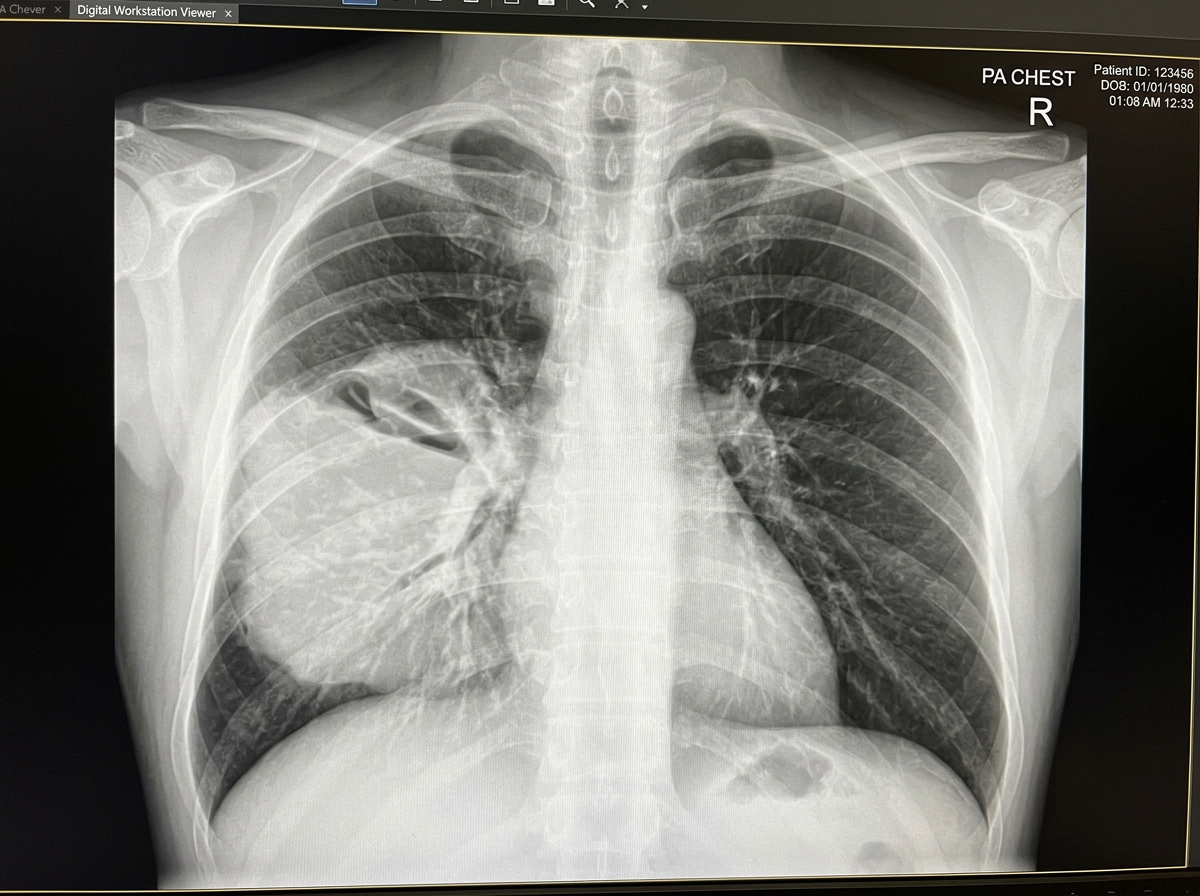
Right Lower Lobe Consolidation Lobar Pneumonia CXR
Posterior-anterior (PA) chest radiograph demonstrating classic dense Right Lower Lobe alveolar consolidation with air bronchograms, pathognomonic for Community-Acquired Lobar Pneumonia (most commonly *Streptococcus pneumoniae*). Note preservation of the right heart border (indicating middle lobe sparing) with obliteration of the posterior costophrenic angle.
HIV Opportunistic Infection Prophylaxis Thresholds
Clinical Matrix
CD4 Count ThresholdTargeted Opportunistic PathogenFirst-Line ChemoprophylaxisKey Clinical Features
CD4 < 200 cells/µLPneumocystis jirovecii Pneumonia (PJP)Trimethoprim-Sulfamethoxazole (TMP-SMX, 1 DS tab daily)Bilateral ground-glass infiltrates, exertional hypoxia, high LDH; add steroids if PaO2 < 70
CD4 < 100 cells/µLToxoplasma gondiiTMP-SMX (prophylaxis against both PJP and Toxo)Multiple ring-enhancing brain lesions with basal ganglia predilection; treat with pyrimethamine-sulfadiazine
CD4 < 50 cells/µLMycobacterium avium Complex (MAC)Azithromycin 1200 mg weekly (only if NOT on ART)High fever, night sweats, diarrhea, hepatosplenomegaly, alkaline phosphatase elevation
Any CD4 with positive PPD (≥ 5 mm)Mycobacterium tuberculosis (latent TB)Isoniazid + Pyridoxine (Vit B6) × 9 mo, or Rifampin × 4 moPrevents peripheral neuropathy caused by B6 depletion
Board Trap — Head CT Prior to Lumbar Puncture Indications
High-Yield Board Trap & Alert
A 42-year-old male presents with fever, severe headache, photophobia, and neck stiffness. Do NOT delay antibiotic administration for a head CT! In suspected meningitis, blood cultures must be drawn and empiric IV antibiotics + dexamethasone administered immediately if CT will delay lumbar puncture. A screening non-contrast head CT prior to LP is only required if specific high-risk signs of elevated intracranial pressure / herniation risk are present: (1) Focal neurologic deficits; (2) New-onset seizures; (3) Papilledema; (4) Altered mental status / depressed GCS; or (5) Severe immunocompromised state.

Chapter 6 Quick-Check Self-Assessment

Test your clinical reasoning before moving to the next chapter
3 Vignettes
Board Vignette #1 Single Best Answer
A 56-year-old male with fever and a new holosystolic murmur has 3 sets of blood cultures positive for Streptococcus gallolyticus (formerly S. bovis). Transesophageal echocardiography reveals an oscillating vegetation on the posterior mitral leaflet. In addition to IV ceftriaxone, what diagnostic procedure is mandatory?
Board Vignette #2 Single Best Answer
A 67-year-old female presents with fever, neck stiffness, and confusion. Lumbar puncture is planned. Which of the following empiric antimicrobial regimens must be ordered for her age group?
Board Vignette #3 Single Best Answer
A 62-year-old female receiving adjuvant chemotherapy for breast cancer presents with fever (101.8°F) and fatigue. Absolute neutrophil count (ANC) is 320 cells/mm3. Physical examination, chest X-ray, and urinalysis reveal no localized source of infection. What is the most appropriate initial empiric antimicrobial therapy?
Estimated study time: 14 min
Chapter 7 • Rheumatology & Clinical Immunology
13 min TOC

Rheumatology, Autoimmunity & Systemic Vasculitis

Rheumatoid Arthritis vs. Osteoarthritis, Systemic Lupus Erythematosus (SLE), Crystal Arthropathies, and Giant Cell Arteritis

Clinical Overview & Board Focus

Rheumatology questions test the clinical differentiation of inflammatory vs. mechanical joint pain, interpretation of synovial fluid analysis, specific autoantibody panels, and time-sensitive treatment of giant cell arteritis to preserve vision.

7.1 Crystal Arthropathies, Systemic Lupus & Giant Cell Arteritis

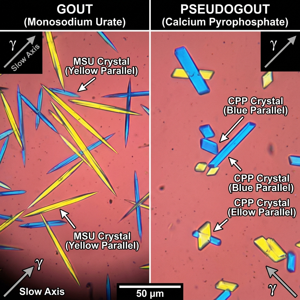
Crystal Arthropathies present with acute, exquisite monoarthritis. Gout results from supersaturation and tissue deposition of Monosodium Urate (MSU) crystals, most commonly in the first metatarsophalangeal joint (podagra). Synovial fluid polarized light microscopy demonstrates Needle-shaped, strongly negatively birefringent crystals (yellow when parallel to the compensator axis, blue when perpendicular). Acute flare treatment: NSAIDs (Indomethacin), oral Colchicine (inhibits microtubule polymerization), or systemic Corticosteroids (first-line if renal disease or CHF contraindicates NSAIDs). Long-term urate-lowering therapy (Allopurinol, Febuxostat; xanthine oxidase inhibitors) targeting serum urate < 6.0 mg/dL is indicated for ≥ 2 flares/year, tophi, or CKD; allopurinol must NEVER be initiated during an acute flare without concomitant anti-inflammatory prophylaxis. Pseudogout (Calcium Pyrophosphate Dihydrate [CPPD] crystal deposition disease) classically affects the knee or wrist, often associated with hemochromatosis, hyperparathyroidism, or hypomagnesemia. Synovial fluid shows Rhomboid-shaped, weakly positively birefringent crystals (blue when parallel), with chondrocalcinosis (cartilage calcification) on radiographs.

Systemic Lupus Erythematosus (SLE) is a systemic autoimmune disease characterized by immune complex deposition affecting reproductive-aged women. Clinical features span the classic SLICC criteria: malar butterfly rash (sparing nasolabial folds), discoid rash, photosensitivity, painless oral ulcers, non-erosive arthritis, serositis (pleuritis, pericarditis), lupus nephritis (diffuse proliferative glomerulonephritis [class IV] is most severe), and hematologic cytopenias. Serologies: Antinuclear Antibodies (ANA) boast > 99% sensitivity (excellent screening test); Anti-double-stranded DNA (anti-dsDNA, correlates with active lupus nephritis and disease flares) and Anti-Smith (anti-Sm) have exceptional specificity (> 99%). All SLE patients should receive Hydroxychloroquine (reduces flare frequency, organ damage, and mortality; requires annual ophthalmologic screening for retinal toxicity).

Giant Cell (Temporal) Arteritis (GCA) is a large-vessel granulomatous vasculitis occurring in patients > 50 years, intimately linked with Polymyalgia Rheumatica (PMR: morning stiffness in bilateral shoulders and hips). GCA presents with new-onset unilateral headache, scalp tenderness, jaw claudication (pain with chewing due to maxillary artery ischemia), and amaurosis fugax (transient vision loss). Erythrocyte sedimentation rate (ESR) is markedly elevated, typically > 50 mm/hr (often > 100 mm/hr). Immediate initiation of high-dose systemic Corticosteroids (oral Prednisone 60 mg daily, or IV Methylprednisolone if vision changes are present) is MANDATORY to prevent permanent irreversible ischemic optic neuropathy (blindness) and must NEVER be delayed while waiting for temporal artery biopsy.

Gout vs Pseudogout Polarized Light Microscopy
Polarized light microscopy comparison: Gout features needle-shaped monosodium urate crystals demonstrating strongly negative birefringence (yellow under parallel polarizing light, blue when perpendicular). Pseudogout (CPPD) features rhomboid calcium pyrophosphate crystals demonstrating weakly positive birefringence (blue when parallel). First-line acute therapy for gout: NSAIDs (Indomethacin), Colchicine, or Intra-articular Steroids.
Synovial Fluid Analysis Differential Matrix
Clinical Matrix
ClassificationWBC Count (/µL)Neutrophils (%)Appearance & ViscosityClassic Clinical Etiologies
Normal< 200< 25%Clear, transparent, high viscosityNormal joint
Non-Inflammatory200 – 2,000< 25%Clear to slightly yellow, high viscosityOsteoarthritis, trauma, osteonecrosis
Inflammatory2,000 – 50,000≥ 50%Translucent to opaque, yellow, low viscosityRheumatoid Arthritis, Gout, Pseudogout, Reactive arthritis, SLE
Septic> 50,000 (often > 100k)≥ 75% (often > 90%)Opaque, purulent, turbid, very low viscosityBacterial Septic Arthritis (Staph aureus, Neisseria gonorrhoeae)
Board Trap — Temporal Artery Biopsy Timing
High-Yield Board Trap & Alert
A 72-year-old female presents with severe right temporal headache, scalp pain when brushing her hair, jaw fatigue when chewing steak, and ESR of 104 mm/hr. The emergency physician schedules a temporal artery biopsy for the following afternoon but holds steroids until the biopsy is performed so it 'won't mask pathology'. THIS IS DANGEROUS MALPRACTICE! High-dose glucocorticoids must be started IMMEDIATELY. Histopathologic evidence of granulomatous vasculitis and giant cells on temporal artery biopsy remains detectable for up to 2 to 4 weeks after initiating corticosteroid therapy.

Chapter 7 Quick-Check Self-Assessment

Test your clinical reasoning before moving to the next chapter
3 Vignettes
Board Vignette #1 Single Best Answer
A 58-year-old male with chronic kidney disease (eGFR 24 mL/min) presents with acute, severe pain, erythema, and swelling of his left knee. Arthrocentesis reveals 28,000 WBCs/µL (65% neutrophils) and rhomboid-shaped, weakly positively birefringent crystals under polarized light microscopy. Knee radiograph reveals punctate radiodense calcification of the articular cartilage (chondrocalcinosis). What is the preferred acute medical management?
Board Vignette #2 Single Best Answer
A 28-year-old female with Systemic Lupus Erythematosus presents for routine follow-up. She is asymptomatic on hydroxychloroquine. Serum labs reveal creatinine 0.9 mg/dL. Urinalysis demonstrates 3+ proteinuria and 20 dysmorphic RBCs per high-power field with occasional RBC casts. Serum complement C3 and C4 are low, and anti-dsDNA titer is markedly elevated. What is the most appropriate next clinical step?
Board Vignette #3 Single Best Answer
A 34-year-old sexually active male presents with acute pain, swelling, and erythema of the right knee. Arthrocentesis yields cloudy synovial fluid with 68,000 WBC/mm3 (88% neutrophils) and gram-negative intracellular diplococci. What is the definitive antimicrobial regimen?
Estimated study time: 13 min