Skip to content
NBOME Emergency Medicine COMAT & Level 2-CE NBME Emergency Medicine Shelf Exam & Step 2 CK

Emergency Medicine COMAT Command Center Emergency Medicine Shelf Exam Command Center

Acute resuscitation, trauma, toxicology, and emergency procedures guide for NBOME Emergency Medicine COMAT and COMLEX Level 2-CE. Ingests 1,850+ acute care cards paired with 21 dedicated clinical emergency protocols.

70 Cards Extracted 2 High-Yield Protocols 3 Osteopathic Rules 4 High-Yield Pearls
Section 01 • Osteopathic Principles

"Don't Miss" COMAT Osteopathic Pearls

High-frequency somatic dysfunctions, viscerosomatics, autonomic reflexes, and treatment rules

Tested heavily on NBOME Shelf

Contraindications to HVLA in Trauma & Emergency

Absolute contraindications: Suspected spinal fractures, acute dislocation, cervical spine instability (Down syndrome, rheumatoid arthritis with transverse atlantal ligament laxity), vertebral artery insufficiency, acute osteomyelitis, and active bone malignancy. Relative: Acute herniated disc, severe osteoporosis, patients on full therapeutic anticoagulation.

Autonomic Stabilization in Shock & Sepsis

In severe hypovolemic, septic, or cardiogenic shock, avoid vigorous lymphatic pumps that can transiently overload the right atrium or dislodge occult thrombi. Instead, utilize gentle paraspinal rib raising and suboccipital release to normalize sympathetic outflow and reduce systemic peripheral vascular resistance.

Appendicitis & Acute Abdomen Chapman Point

Classic acute appendicitis Chapman reflex point: Located on the tip of the right 12th rib anteriorly; between T11 and T12 transverse processes on the right posteriorly. Associated with marked right psoas hypertonicity and right pelvic tilt.

Section 01 • Clinical Foundations

"Don't Miss" Emergency Medicine Clinical Pearls & Shelf Traps

High-frequency diagnostic pitfalls, gold-standard criteria, and next-best-step clinical rules

Tested heavily on NBME Shelf & Step 2 CK

Undifferentiated Hypotension & RUSH Protocol Windows

Rapid Ultrasound for Shock and Hypotension (RUSH) evaluates the Pump, Tank, and Pipes. 1. The Pump: Parasternal long/short-axis and apical 4-chamber views assess contractility, pericardial effusion/tamponade (diastolic RV collapse), and acute RV strain (McConnell sign in massive PE). 2. The Tank: IVC collapsibility (< 50% collapse with inspiration suggests volume overload/tamponade; > 50% collapse suggests hypovolemia), FAST views for intra-abdominal free fluid, and lung ultrasound for pneumothorax (absent lung sliding, stratosphere sign). 3. The Pipes: Abdominal aorta diameter (> 3 cm = aneurysm; > 5 cm = rupture risk) and bilateral lower extremity compression ultrasound for DVT.

Massive Transfusion Protocol (MTP) & Lethal Triad Prevention

Initiate MTP for traumatic hemorrhagic shock (Assessment of Blood Consumption [ABC] score >= 2: penetrating mechanism, SBP <= 90 mmHg, HR >= 120 bpm, positive FAST). Transfuse balanced 1:1:1 ratio of Packed Red Blood Cells (PRBCs), Fresh Frozen Plasma (FFP), and Platelets. Administer Tranexamic Acid (TXA 1 g IV over 10 min, then 1 g over 8 hr) within 3 hours of injury. Aggressively combat the Lethal Triad: Hypothermia (warmed fluids, external warming), Acidosis (restore perfusion), and Coagulopathy (monitor calcium: replace ionized Ca2+ due to citrate toxicity).

Acute Coronary Syndrome Rule-Out & High-Sensitivity Troponin

High-sensitivity cardiac troponin (hs-cTn) protocols allow rapid 0- and 1- or 2-hour delta evaluation. The HEART score (History, ECG, Age, Risk factors, Troponin) stratifies chest pain risk: scores 0–3 permit safe outpatient discharge with rapid follow-up (< 1% 30-day major adverse cardiac event [MACE]); scores 4–6 require inpatient telemetry and observation; scores >= 7 warrant early invasive coronary angiography. Critical rule: Beta-blockers are strictly contraindicated in cocaine-induced acute chest pain due to unopposed alpha-1 vasoconstriction precipitating coronary spasm and malignant hypertension.

Status Epilepticus Stepwise Pharmacological Escalation

Phase 1 (0–5 min): Stabilize ABCs, check fingerstick glucose, give IV Lorazepam 4 mg (or IM Midazolam 10 mg if no IV access). May repeat once at 5–10 min. Phase 2 (10–20 min): If seizure continues, administer IV second-line antiepileptic: Levetiracetam (Keppra 60 mg/kg, max 4500 mg), Fosphenytoin (20 mg PE/kg), or Sodium Valproate (40 mg/kg). Phase 3 (20–40 min, Refractory Status): Proceed immediately to endotracheal intubation and continuous IV anesthetic infusion: Propofol (titrated to burst suppression on continuous EEG), Midazolam infusion, or Ketamine.

Section 02 • Clinical Algorithms

Core Clinical Protocols & Diagnostic Trees

Step-by-step first-line management pathways, diagnostic thresholds, and pharmacological escalation

2 Diagnostic Algorithms

ACLS Cardiac Arrest & Shockable Rhythms Algorithm

  1. 1. Verify cardiac arrest: Check pulse/breathing (< 10 seconds). Call for code team and defibrillator; begin high-quality CPR (100-120 bpm, 2-2.4 in depth, full recoil).
  2. 2. Rhythm check: Ventricular Fibrillation (VF) or Pulseless Ventricular Tachycardia (pVT) -> Immediate Defibrillation (120-200 J biphasic).
  3. 3. Resume CPR immediately for 2 minutes without checking pulse post-shock; obtain IV/IO access.
  4. 4. Shock 2: Deliver shock, resume CPR -> Epinephrine 1 mg IV/IO every 3-5 minutes.
  5. 5. Shock 3: Deliver shock, resume CPR -> Amiodarone 300 mg IV bolus (or Lidocaine 1-1.5 mg/kg); second dose 150 mg after next cycle.
  6. 6. Search for reversible causes (H's & T's): Hypovolemia, Hypoxia, Hydrogen ion (acidosis), Hypo/hyperkalemia, Hypothermia; Tension pneumothorax, Tamponade, Toxins, Thrombosis (pulmonary/coronary).

Primary Trauma Survey (ABCDE) Protocol

  1. 1. Airway with cervical spine protection: Assess patency, clear secretions; maintain manual in-line cervical stabilization; perform RSI if GCS <= 8.
  2. 2. Breathing and ventilation: Inspect chest rise, auscultate bilaterally. Rule out immediate threats: Tension pneumothorax (needle decompression 2nd ICS MCL or 5th ICS AAL followed by chest tube), open pneumothorax (3-sided occlusive dressing), flail chest, massive hemothorax.
  3. 3. Circulation with hemorrhage control: Direct manual pressure on external bleeding; apply tourniquets to extremities; assess pulses, capillary refill, skin color; establish two large-bore 16G peripheral IVs or IO access. Initiate balanced massive transfusion protocol (1:1:1 PRBC, FFP, platelets).
  4. 4. Disability (Neurologic status): GCS, pupillary symmetry, gross motor movement, point-of-care blood glucose.
  5. 5. Exposure and environmental control: Completely undress patient, inspect entire skin; warm blanket application to prevent lethal triad (hypothermia, coagulopathy, acidosis).
Section 03 • Clinical Chapters & Active Recall

High-Yield Clerkship Review by System

Read structured textbook-style disease summaries or test yourself with active-recall flashcards across Emergency Medicine clinical systems.

Showing 7 of 7 continuous textbook chapters
Chapter 1 • Resuscitation, Shock & ACLS
14 min TOC

Resuscitation, Hemodynamic Shock & ACLS

Undifferentiated Hypotension, Shock Profiles, Cardiac Arrest, and Vasoactive Resuscitation

Clinical Overview & Board Focus

The initial stabilization of the critically ill patient with undifferentiated hypotension requires rapid identification of the underlying shock etiology through targeted physical examination, invasive hemodynamics or point-of-care ultrasound (RUSH exam), and aggressive restoration of tissue perfusion. Board questions demand mastery of the four primary shock classes, ACLS dysrhythmia pathways, and appropriate selection of inotropes and vasopressors.

1.1 Pathophysiologic Classification of Shock & Hemodynamic Profiles

Shock is defined at the cellular level as acute circulatory failure leading to inadequate tissue perfusion, resulting in cellular hypoxia, anaerobic metabolism, and systemic lactic acidosis. Clinically, shock is categorized into four distinct pathophysiological phenotypes: hypovolemic, cardiogenic, obstructive, and distributive. In hypovolemic shock (hemorrhage, severe dehydration, third-spacing), decreased circulating plasma volume causes marked reductions in cardiac output (CO) and pulmonary capillary wedge pressure (PCWP/preload), triggering compensatory peripheral vasoconstriction that manifests as markedly elevated systemic vascular resistance (SVR). Patients present with cold, clammy extremities, collapsed jugular veins, and tachycardia.

Cardiogenic shock occurs due to primary pump failure, most frequently secondary to massive acute myocardial infarction, severe acute valvular regurgitation (e.g., papillary muscle rupture), or decompensated cardiomyopathy. Hemodynamically, cardiac contractility is profoundly impaired, leading to severely decreased cardiac output, elevated ventricular end-diastolic pressures (high PCWP), and compensatory elevated SVR. On physical exam, patients present with pulmonary edema (crackles), elevated jugular venous distention (JVD), an S3 gallop, and cool extremities. First-line medical support includes inotropes (dobutamine, milrinone) to enhance forward flow, vasopressors (norepinephrine) if mean arterial pressure (MAP) is < 65 mmHg, and prompt mechanical circulatory support (intra-aortic balloon pump [IABP] or Impella) with emergency catheterization.

Obstructive shock is characterized by physical impedance to cardiac chamber filling or right ventricular outflow. The classic etiologies include massive pulmonary embolism (RV strain, elevated CVP, low PCWP, low CO), cardiac tamponade (impaired diastolic filling of all chambers with equalization of diastolic pressures and pulsus paradoxus > 10 mmHg), and tension pneumothorax (mediastinal shift kinking the inferior vena cava). In contrast, distributive shock—predominantly septic shock, anaphylaxis, and neurogenic shock—is marked by severe pathological systemic vasodilation. Here, SVR is profoundly decreased, and in early 'warm shock', cardiac output is elevated or normal with warm, hyperemic extremities and bounding pulses. Neurogenic shock represents a unique distributive state caused by high cervical or thoracic spinal cord transection (above T6), disrupting descending sympathetic tone and resulting in the classic hemodynamic triad of hypotension, relative bradycardia, and warm dry skin without compensatory vasoconstriction.
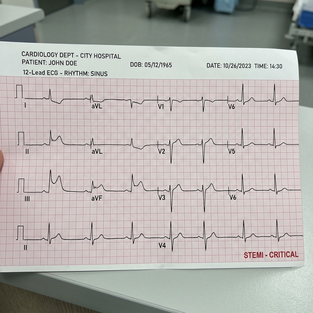
Inferior STEMI ECG Leads II, III, aVF
Classic Inferior STEMI demonstrating prominent ST-segment elevation in leads II, III, and aVF with reciprocal ST depression in lead aVL. Sourced from right coronary artery (RCA) occlusion. *Critical rule:* Always obtain right-sided lead V4R before administering nitroglycerin—preload reduction in right ventricular infarction precipitates catastrophic refractory hypotension.
Sepsis-3 Resuscitation & Vasoactive Pressor Escalation Matrix
Clinical Matrix
Phase / AgentClinical MechanismHemodynamic TargetHigh-Yield Board Caveat
Crystalloid Bolus (Hour-1)Balanced crystalloid 30 mL/kg within 3 hrMAP ≥ 65 mmHg; lactate clearanceAvoid 0.9% normal saline boluses in large volumes due to hyperchloremic metabolic acidosis.
Norepinephrine (Levophed)Potent alpha-1 vasoconstriction > beta-1 inotropyFirst-line vasopressor of choice in septic & cardiogenic shockTitrate rapidly via central venous access; maintains renal/splanchnic perfusion.
Vasopressin (0.03 units/min)V1 vascular smooth muscle constriction (non-adrenergic)Second-line catecholamine-sparing agentDo NOT titrate; fixed dose added when norepinephrine requirements exceed 0.25 mcg/kg/min.
Epinephrine (Adrenaline)Potent beta-1, beta-2 (low dose) and alpha-1 (high dose)First-line in anaphylactic shock; 2nd/3rd line in refractory septic shockProduces transient hyperlactatemia via aerobic skeletal muscle glycolysis (type B hyperlactatemia).
DobutamineBeta-1 inotrope > beta-2 vasodilatorAdded in persistent hypoperfusion despite adequate volume and MAPContraindicated as monotherapy in hypotension; causes reflex vasodilation.
Cardiopulmonary Autonomics & Thoracic Inlet Release
COMLEX & OPP Board Integration
Sympathetic innervation to the myocardium arises from T1–T5 (right sympathetic chain supplies SA node -> SVT/sinus tachycardia; left sympathetic chain supplies AV node -> ventricular dysrhythmias). Parasympathetic control via Vagus nerve (CN X) slows nodal conduction. During septic and hemodynamic shock, opening the Thoracic Inlet (Sibson's fascia) and performing gentle rib raising down-regulates hypersympathetic constriction and dramatically enhances terminal lymphatic drainage through the thoracic duct into the left subclavian vein.

1.2 Advanced Cardiac Life Support (ACLS) & Rapid Sequence Intubation (RSI)

In cardiac arrest, rhythm analysis partitions cases into shockable rhythms (Ventricular Fibrillation [VF] and Pulseless Ventricular Tachycardia [pVT]) and non-shockable rhythms (Pulseless Electrical Activity [PEA] and Asystole). For VF/pVT, immediate defibrillation (unsynchronized high-energy shock) is the single most critical determinant of survival, followed immediately by 2 minutes of uninterrupted, high-quality chest compressions (100–120 bpm, 2–2.4 inches depth, allowing complete chest recoil). Epinephrine 1 mg IV/IO is administered every 3 to 5 minutes, beginning after the second defibrillation cycle. Amiodarone (300 mg bolus, followed by a 150 mg second dose) or Lidocaine (1–1.5 mg/kg) is administered after the third shock for refractory VF/pVT.

For non-shockable rhythms (PEA/Asystole), defibrillation is strictly contraindicated. CPR and immediate administration of Epinephrine 1 mg IV/IO must occur without delay. The clinician must relentlessly search for and treat reversible underlying causes using the classic 5 H's and 5 T's: Hypovolemia, Hypoxia, Hydrogen ion (acidosis), Hypo-/Hyperkalemia, Hypothermia; Tension pneumothorax, Tamponade (cardiac), Toxins, Thrombosis (pulmonary PE), and Thrombosis (coronary ACS). Return of spontaneous circulation (ROSC) is evidenced by an abrupt, sustained increase in end-tidal CO2 (PETCO2 ≥ 40 mmHg) or arterial pressure waveforms.

Emergency airway management in the resuscitation bay hinges upon Rapid Sequence Intubation (RSI)—the virtually simultaneous administration of a potent induction agent and a neuromuscular blocking agent to facilitate endotracheal tube placement while minimizing aspiration risk. Etomidate (0.3 mg/kg) is widely favored for its exceptional hemodynamic neutrality; however, its transient suppression of 11-beta-hydroxylase must be considered in severe septic shock. Ketamine (1.5–2 mg/kg) is ideal for hypotensive trauma and status asthmaticus due to its catecholamine-releasing sympathomimetic properties and intrinsic bronchodilation. Succinylcholine (1.5 mg/kg) is a depolarizing paralytic with rapid 45-second onset, but is strictly contraindicated in hyperkalemia, severe crush injuries > 48 hours old, extensive burns > 48 hours old, chronic denervating diseases (ALS, MS), and malignant hyperthermia susceptibility. In such settings, high-dose Rocuronium (1.2 mg/kg) is the paralytic of choice, which can be rapidly reversed with Sugammadex if needed.

Board Trap — Succinylcholine Hyperkalemia Lethality
High-Yield Board Trap & Alert
Succinylcholine typically causes a transient, predictable increase in serum potassium of 0.5 to 1.0 mEq/L. However, in patients with denervation injuries (spinal cord injury, stroke > 72 hours), extensive burns, or severe crush injuries, massive upregulation of post-synaptic nicotinic acetylcholine receptors occurs throughout the sarcolemma. Administering succinylcholine to these patients can cause catastrophic efflux of intracellular potassium, precipitating fatal cardiac arrest from hyperkalemic sinusoidal arrhythmias within minutes. Always select Rocuronium (1.2 mg/kg) when burn, crush, or denervation history is present.

Chapter 1 Quick-Check Self-Assessment

Test your clinical reasoning before moving to the next chapter
3 Vignettes
Board Vignette #1 Single Best Answer
A 72-year-old female presents in acute shock. Invasive hemodynamic monitoring reveals: CVP 18 mmHg (elevated), Cardiac Output 2.1 L/min (profoundly decreased), PCWP 26 mmHg (elevated), and SVR 1,900 dynes·sec/cm⁵ (elevated). What is the underlying shock phenotype?
Board Vignette #2 Single Best Answer
A 26-year-old male is brought to the trauma bay following a high-speed motorcycle collision. He suffered an acute fracture-dislocation at C5 with complete spinal cord transection. Blood pressure is 82/46 mmHg, heart rate is 48 bpm, respirations are 18/min, and temperature is 36.8°C. His skin is warm, flushed, and dry with brisk capillary refill. Neurological exam reveals flaccid quadriplegia and loss of sensation below the clavicles. Intravenous crystalloid bolus produces minimal change in hemodynamics. Which of the following is the most appropriate next step?
Board Vignette #3 Single Best Answer
A 64-year-old male with acute inferior STEMI receives sublingual nitroglycerin and suddenly becomes unresponsive with BP 72/40 mmHg and HR 52 bpm. Physical exam shows clear lung sounds bilaterally and marked jugular venous distension. What is the immediate next step in management?
Estimated study time: 14 min
Chapter 2 • Trauma & Musculoskeletal
14 min TOC

Trauma Resuscitation, Primary Survey & Orthopedic Emergencies

ATLS Protocols, Massive Transfusion, Open Fractures, Compartment Syndrome, and Spine Clearance

Clinical Overview & Board Focus

Trauma is governed by strict adherence to Advanced Trauma Life Support (ATLS) algorithms: Airway with cervical spine protection, Breathing and ventilation, Circulation with hemorrhage control, Disability (neurological evaluation), and Exposure/environmental control. Board exams heavily test the prioritization of life-saving interventions, FAST ultrasound interpretation, and the immediate recognition of time-sensitive orthopedic catastrophes.

2.1 ATLS Primary Survey & Massive Transfusion Protocol (MTP)

The primary survey begins with Airway assessment while maintaining rigid in-line cervical spine stabilization. Any patient with a Glasgow Coma Scale (GCS) score ≤ 8, severe maxillofacial trauma, expanding neck hematoma, or severe inhalation burn requires definitive airway placement via endotracheal intubation. Breathing assessment entails immediate detection of tension pneumothorax (unilateral absent breath sounds, tracheal deviation, hemodynamic collapse), which mandates immediate needle decompression at the 2nd intercostal space midclavicular line or 4th/5th intercostal space anterior axillary line, immediately followed by tube thoracostomy (chest tube). Flail chest, defined by ≥ 2 fractures in ≥ 2 consecutive ribs, creates paradoxical chest wall motion and severe underlying pulmonary contusion; definitive therapy emphasizes early analgesia (epidural or intercostal nerve blocks) and positive pressure ventilation if respiratory failure ensues.

Circulation centers on rapid hemorrhage control and resuscitation. In hemorrhagic shock, crystalloid boluses are strictly limited to avoid hypothermia, hemodilution, and worsening trauma-induced coagulopathy ('the lethal triad': hypothermia, acidosis, and coagulopathy). The standard of care is early activation of the Massive Transfusion Protocol (MTP), delivering packed red blood cells, fresh frozen plasma, and platelets in a balanced 1:1:1 ratio. Tranexamic acid (TXA, 1 g IV over 10 minutes followed by 1 g infusion over 8 hours) must be administered within 3 hours of injury to significantly reduce all-cause mortality from trauma hemorrhage.

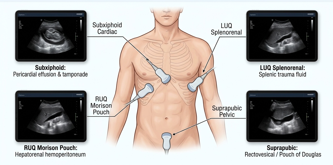
The Focused Assessment with Sonography for Trauma (FAST) exam rapidly screens four acoustic windows for pathological free fluid: the hepatorenal space (Morison's pouch), the splenorenal recess, the pelvis/rectovesical or rectouterine pouch, and the subxiphoid pericardial window (extended FAST adds bilateral anterior chest views for pneumothorax). In an unstable trauma patient, a positive FAST indicates hemoperitoneum and mandates immediate emergent exploratory laparotomy without waiting for CT imaging. In a hemodynamically stable patient, a positive FAST warrants contrast-enhanced abdominopelvic CT to grade solid organ injury (spleen, liver, kidney) and guide non-operative vs. interventional angiographic embolization.

FAST Ultrasound 4 Acoustic Windows Diagram
The FAST Exam (Focused Assessment with Sonography for Trauma) evaluating 4 anatomical acoustic windows: 1) Morison's pouch (hepatorenal space), 2) Splenorenal recess, 3) Suprapubic pelvic cavity (rectovesical / pouch of Douglas), and 4) Subxiphoid pericardial window. In an unstable trauma patient, free fluid in Morison's pouch mandates immediate exploratory laparotomy without CT scanning.
Trauma Massive Transfusion Protocol (MTP) & Lethal Triad Prevention
Clinical Matrix
Component / TargetAdministration RatioClinical RationaleBoard Trap to Avoid
Balanced Blood Products1:1:1 Ratio (pRBCs : FFP : Platelets)Prevents dilutional coagulopathy from crystalloid over-resuscitationDo NOT wait for lab results (PT/INR/fibrinogen) before activating MTP in hemorrhagic shock.
Tranexamic Acid (TXA)1 g IV over 10 min, then 1 g over 8 hrAntifibrinolytic (inhibits plasminogen activation); CRASH-2 trial mortality benefitMust be given within 3 hours of injury; administration > 3 hours increases mortality.
Calcium Chloride (1 g IV)1 amp IV for every 4 units of blood productsReplaces ionized calcium bound by citrate anticoagulant in blood bagsCitrate toxicity causes severe hypocalcemia -> refractory hypotension and prolonged QTc.
Active RewarmingForced-air warming blankets & IV fluid warmersHypothermia (<35°C) halts coagulation cascade enzyme kineticsEvery 1°C drop in core temperature decreases coagulation factor activity by 10%.
COMLEX Clinical Integration — Rib Kinematics & Pulmonary Complications in Chest Trauma
COMLEX & OPP Board Integration
Rib fractures and thoracic wall trauma cause severe respiratory splinting, hypoventilation, and rapid development of atelectasis and secondary pneumonia. Knowledge of rib motion kinematics is paramount: Ribs 1–5 move predominantly in a 'pump-handle' motion (increasing anteroposterior diameter), Ribs 6–10 exhibit 'bucket-handle' motion (increasing transverse diameter), and Ribs 11–12 move in 'caliper' motion. Osteopathic intervention should avoid direct HVLA to fractured segments. Gentle rib raising and myofascial release of the thoracic inlet, scalenes, and diaphragm relieve respiratory restriction, improve lymphatic drainage via the thoracic duct, and prevent post-traumatic atelectasis.

Chapter 2 Quick-Check Self-Assessment

Test your clinical reasoning before moving to the next chapter
3 Vignettes
Board Vignette #1 Single Best Answer
A 28-year-old unrestrained driver arrives at the trauma bay following a high-speed collision. He is cyanotic and tachypneic (RR 36/min). Vitals: BP 70/40 mmHg, HR 138 bpm, SpO2 78%. Physical exam reveals absent breath sounds over the entire right hemithorax, hyperresonance to percussion on the right, distended neck veins, and the trachea deviated to the left. What is the immediate life-saving action?
Board Vignette #2 Single Best Answer
A 32-year-old unrestrained driver is involved in a high-speed motor vehicle collision. In the trauma bay, he is hemodynamically stable. Pelvic examination reveals gross instability with pelvic rock. Physical exam reveals blood at the urethral meatus, perineal ecchymosis, and a high-riding, non-palpable prostate on digital rectal examination. What is the most appropriate next diagnostic step?
Board Vignette #3 Single Best Answer
A 28-year-old restrained driver presents following a high-speed motor vehicle collision. Blood pressure is 78/48 mmHg and heart rate is 134 bpm. FAST exam reveals free fluid in the hepatorenal recess (Morison's pouch). What is the definitive management?
Estimated study time: 14 min
Chapter 3 • Toxicology & Poisoning
13 min TOC

Emergency Toxicology, Toxidromes & Antidotes

Acetaminophen Toxicity, Toxic Alcohols, Anticholinergic vs. Sympathomimetic Toxidromes, and Chelators

Clinical Overview & Board Focus

Emergency toxicology requires pattern recognition of classic clinical toxidromes, rapid calculation of the anion gap and osmolal gap, and prompt administration of targeted antidotes. Board questions frequently present patients with altered mental status, abnormal pupil size, and vital sign extremes, demanding rapid pinpointing of the offending drug or poison.

3.1 Clinical Toxidromes & Pattern Recognition

Toxidromes represent clinical symptom constellations that point toward specific classes of chemical exposures. The Anticholinergic toxidrome (atropine, diphenhydramine, tricyclic antidepressants, scopolamine) is remembered by the classic medical aphorism: 'Blind as a bat (mydriasis, loss of accommodation), mad as a hatter (delirium, hallucinations), red as a beet (cutaneous flushing), hot as a hare (hyperthermia due to anhidrosis), dry as a bone (dry skin and mucous membranes), and full as a flask (urinary retention and decreased bowel sounds)'. In contrast, the Sympathomimetic toxidrome (cocaine, amphetamines, MDMA, cathinones) presents nearly identically with hyperthermia, mydriasis, hypertension, and tachycardia, but is crucially distinguished by diaphoresis (sweating) and hyperactive bowel sounds.

The Cholinergic toxidrome (organophosphate and carbamate insecticides, chemical nerve agents like sarin) inhibits acetylcholinesterase, producing massive acetylcholine accumulation at muscarinic and nicotinic receptors. Muscarinic overstimulation is remembered by DUMBELS: Defecation, Urination, Miosis, Bronchorrhea/Bradycardia/Bronchospasm (the 'killer Bs' causing asphyxiation), Emesis, Lacrimation, and Salivation. Nicotinic stimulation causes muscle fasciculations, weakness, and diaphragmatic paralysis. Immediate treatment involves high-dose Atropine (titrated to drying of respiratory secretions) and Pralidoxime (2-PAM, to regenerate active acetylcholinesterase before irreversible covalent aging occurs).

The Opioid toxidrome presents with the classic triad of central nervous system depression, miosis (pinpoint pupils), and respiratory depression (hypoventilation, respiratory acidosis). Immediate airway support and titrated Naloxone (IV, IM, or intranasal) reverses respiratory arrest without precipitating severe withdrawal agitation. Sedative-hypnotic toxicity (benzodiazepines, barbiturates) mimics opioid overdose with CNS depression and hypoventilation, but characteristically features normal pupil size and normal vitals in pure benzodiazepine ingestions; Flumazenil should generally be avoided in chronic users due to the risk of refractory withdrawal seizures.

Emergency Toxicology Antidotes & Mechanism Master Matrix
Clinical Matrix
Toxic Agent / PoisonPathognomonic ToxidromeFirst-Line AntidoteAntidote Mechanism of Action
Acetaminophen (APAP)Asymptomatic early -> AST/ALT > 1,000 U/LN-acetylcysteine (NAC)Replenishes hepatic glutathione; conjugates toxic NAPQI metabolite.
Cyanide (House fire / Nitroprusside)Almond odor, lactic acidosis, bright red venous bloodHydroxocobalaminBinds cyanide to form nontoxic cyanocobalamin (Vitamin B12), excreted in urine.
Carbon Monoxide (CO)Cherry-red lips (rare), headache, carboxyhemoglobin100% Normobaric O2 (Hyperbaric if severe)Displaces CO from hemoglobin (half-life drops from 300m to 90m).
Organophosphates / Nerve AgentsDUMBBELLS cholinergic excess (miosis, bradycardia, wet)Atropine THEN Pralidoxime (2-PAM)Atropine blocks muscarinic receptors; 2-PAM regenerates acetylcholinesterase before aging.
Beta-Blocker ToxicityBradycardia, hypotension, hypoglycemia, cardiogenic shockIV Glucagon (plus High-Dose Insulin)Stimulates adenylate cyclase via non-adrenergic receptors -> increases intracellular cAMP.
Ethylene Glycol / MethanolHigh osmolar gap -> High anion gap metabolic acidosisFomepizole (4-Methylpyrazole)Competitive antagonist of alcohol dehydrogenase; prevents toxic oxalic/formic acid formation.
Board Trap — Pulse Oximetry in Carbon Monoxide & Cyanide
High-Yield Board Trap & Alert
Standard dual-wavelength pulse oximeters CANNOT differentiate between oxyhemoglobin and carboxyhemoglobin because both absorb light at 660 nm identically. Consequently, a patient with lethal carbon monoxide poisoning (carboxyhemoglobin 40%) will falsely display an SpO2 of 99–100%! Arterial or venous blood gas with dedicated co-oximetry is mandatory to measure carboxyhemoglobin. Similarly, in cyanide toxicity, oxygen saturation is normal, but severe uncoupling of cellular oxidative phosphorylation produces massive lactic acidosis and bright red venous blood (elevated central venous oxygen saturation).

Chapter 3 Quick-Check Self-Assessment

Test your clinical reasoning before moving to the next chapter
3 Vignettes
Board Vignette #1 Single Best Answer
A 22-year-old female presents after an intentional overdose of an unknown medication. She is agitated, confused, and hallucinating. Vitals: BP 94/56 mmHg, HR 135 bpm, RR 18/min, Temp 38.8°C (101.8°F). Pupils are 7 mm and sluggishly reactive. Her skin is flushed and warm, axillae are dry, and bowel sounds are absent. ECG reveals a regular sinus tachycardia with a QRS duration of 138 ms and a prominent terminal R wave in lead aVR. What is the definitive initial pharmacotherapy?
Board Vignette #2 Single Best Answer
A 48-year-old male with chronic alcohol use disorder is brought to the ED by EMS after being found unresponsive in an abandoned warehouse next to empty bottles of commercial automotive antifreeze. Vital signs: BP 136/82 mmHg, HR 108 bpm, RR 28 bpm (deep, rapid Kussmaul breathing). Arterial blood gas shows pH 7.12, pCO2 20 mmHg, HCO3 6 mEq/L. Serum sodium 140, potassium 4.2, chloride 100, bicarbonate 6, BUN 34, Cr 2.8 mg/dL. Urinalysis reveals envelope-shaped calcium oxalate crystals. Which of the following is the specific antidote of choice?
Board Vignette #3 Single Best Answer
A firefighter is rescued from an enclosed structural blaze. He presents with severe confusion, headache, BP 90/60 mmHg, HR 118 bpm, and arterial blood gas showing severe lactic acidosis (lactate 12 mmol/L) with bright red venous blood. Pulse oximetry is 99%. What is the most appropriate first-line antidote?
Estimated study time: 13 min
Chapter 4 • Pulmonary & Airway Emergencies
13 min TOC

Acute Pulmonary Emergencies & Respiratory Failure

Massive Pulmonary Embolism, Acute Exacerbation of Asthma & COPD, and Acute Respiratory Distress Syndrome (ARDS)

Clinical Overview & Board Focus

Acute respiratory emergencies range from acute airway obstruction and bronchospastic crises to life-threatening pulmonary vascular occlusion. Board examination questions focus heavily on risk stratification (Wells criteria, PERC rule), selection of appropriate diagnostic imaging, non-invasive positive pressure ventilation (NIPPV) indications and contraindications, and ventilator lung-protective strategies.

4.1 Pulmonary Embolism: Risk Stratification & Acute Management

Venous thromboembolism (VTE) presenting as acute pulmonary embolism (PE) is a major cause of preventable emergency mortality. Evaluation begins with pre-test probability scoring using the Wells Criteria. In patients with low clinical probability (Wells score < 2), the Pulmonary Embolism Rule-out Criteria (PERC: age < 50, HR < 100, SpO2 ≥ 95%, no prior DVT/PE, no recent surgery/trauma within 4 weeks, no hemoptysis, no estrogen use, no unilateral leg swelling) can rule out PE without further diagnostic testing. If any PERC criterion is positive or pretest probability is moderate, a high-sensitivity D-dimer assay is ordered. A negative D-dimer safely excludes PE; an elevated D-dimer warrants definitive diagnostic imaging with CT Pulmonary Angiography (CTPA). If renal insufficiency (GFR < 30) or severe IV contrast allergy precludes CTPA, a Ventilation-Perfusion (V/Q) scan is the gold standard alternative.

PE is classified into three clinical prognostic categories: Massive, Submassive, and Non-massive (low-risk). Massive PE is defined by sustained hypotension (systolic BP < 90 mmHg for > 15 minutes, or requiring vasopressors) due to acute right ventricular afterload mismatch and failure. Submassive PE features normal systemic blood pressure, but exhibits objective evidence of RV dysfunction (elevated troponin, elevated BNP, RV dilation or McConnell's sign on echocardiography, or RV/LV ratio > 0.9 on CTPA). Non-massive PE exhibits neither hypotension nor RV strain.

Treatment for hemodynamically stable PE involves immediate therapeutic anticoagulation with subcutaneous low-molecular-weight heparin (LMWH, Enoxaparin 1 mg/kg BID), Fondaparinux, or direct oral anticoagulants (DOACs like Apixaban or Rivaroxaban). For massive PE with refractory shock or cardiac arrest, immediate systemic thrombolysis with tissue plasminogen activator (Alteplase 100 mg IV over 2 hours) is indicated unless absolute contraindications exist (prior intracranial hemorrhage, known cerebrovascular malformation, ischemic stroke within 3 months, active internal bleeding). If thrombolysis is contraindicated or fails, emergent catheter-directed mechanical thrombectomy or surgical pulmonary embolectomy is indicated.

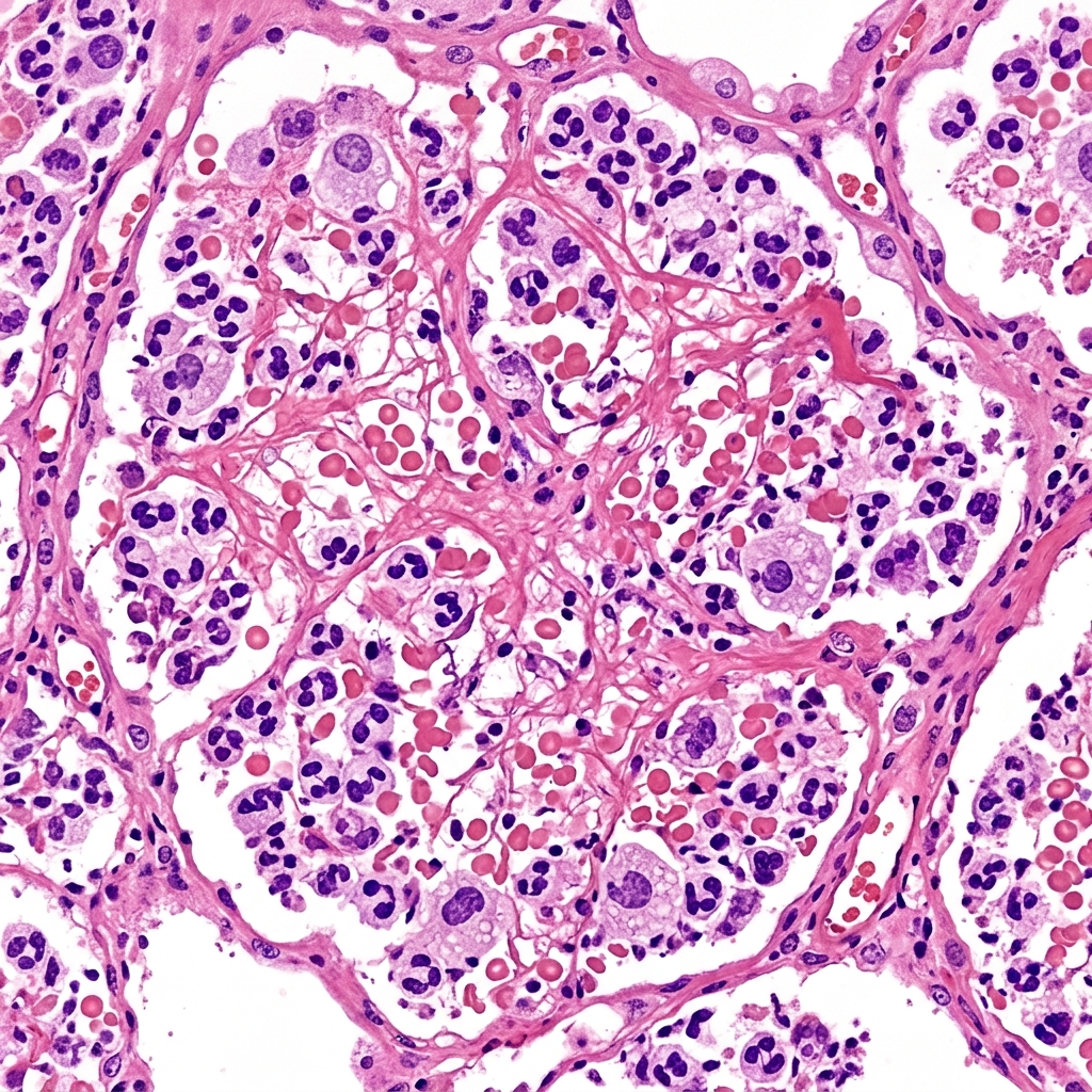
Lobar Pneumonia Consolidation
Histopathology of acute Lobar Pneumonia demonstrating diffuse alveolar exudate rich in neutrophils and fibrin (red/gray hepatization phase). In acute respiratory distress, consolidation produces bronchial breath sounds, dullness to percussion, and increased tactile fremitus.
Acute Respiratory Distress Syndromes & Bronchospasm Matrix
Clinical Matrix
EntityPhysical & ABG FeaturesFirst-Line Medical TherapyVentilator / Airway Considerations
Acute Asthma ExacerbationWheezing, tachypnea, pulsus paradoxus; normal or elevated PaCO2 heralds impending arrestInhaled Albuterol + Ipratropium, IV/PO Methylprednisolone, IV Magnesium sulfate (2 g)Ketamine induction for RSI; allow permissive hypercapnia; low respiratory rate to prevent breath-stacking
Acute COPD ExacerbationIncreased sputum volume, purulence, and dyspnea; respiratory acidosisInhaled bronchodilators, systemic glucocorticoids (Prednisone 40 mg × 5 days), targeted antibioticsEarly trial of BiPAP (NIPPV) reduces intubation and mortality; intubate if encephalopathic or pH < 7.25
ARDSBilateral alveolar infiltrates not explained by heart failure, PaO2/FiO2 ≥ 300, acute onsetTreat underlying cause (sepsis, pancreatitis, trauma); strict fluid restrictionLow tidal volume ventilation (6 mL/kg ideal body weight), plateau pressure < 30 cmH2O, prone positioning
COMLEX Clinical Integration — Thoracic Lymphatic Pump in Pulmonary Consolidation
COMLEX & OPP Board Integration
In acute bronchospastic conditions and pulmonary infections, the thoracic inlet (Sibson's fascia) and diaphragm function as critical mechanical bottlenecks for lymphatic and venous drainage. Osteopathic manipulative treatment should first release the thoracic inlet (first rib, clavicle, T1) to open terminal lymphatic drainage into the left and right subclavian veins. Gentle doming of the abdominal diaphragm and rib raising along T1–T6 normalize sympathetic bronchodilation and reduce interstitial congestion. The thoracic lymphatic pump (Miller technique) should be avoided in acute severe asthma crises with extreme air-trapping, but is highly effective once acute bronchospasm has stabilized.

Chapter 4 Quick-Check Self-Assessment

Test your clinical reasoning before moving to the next chapter
3 Vignettes
Board Vignette #1 Single Best Answer
A 54-year-old male 10 days post-hip arthroplasty presents with sudden severe dyspnea and syncope. Vitals: BP 78/48 mmHg, HR 124 bpm, RR 30/min, SpO2 84% on room air. Bedside echocardiogram reveals acute right ventricular dilatation and McConnell's sign. He has no prior history of stroke or bleeding. What is the most appropriate immediate medical intervention?
Board Vignette #2 Single Best Answer
A 58-year-old female who underwent a total knee arthroplasty 10 days ago presents with acute severe shortness of breath and syncope. On exam, she is diaphoretic, BP is 78/44 mmHg, HR 132 bpm, RR 34/min, and O2 saturation is 84% on a non-rebreather mask. Bedside echocardiogram shows acute right ventricular dilation, McConnell sign (RV free wall hypokinesis with apical sparing), and a flattened interventricular septum bowing into the left ventricle. Following a 500 mL saline bolus, BP remains 80/46 mmHg. What is the most appropriate immediate definitive intervention?
Board Vignette #3 Single Best Answer
A 55-year-old female presents with sudden-onset dyspnea, pleuritic chest pain, and hemoptysis 5 days following right total knee arthroplasty. Oxygen saturation is 88% on room air, HR is 112 bpm, and BP is 128/82 mmHg. What is the diagnostic test of choice?
Estimated study time: 13 min
Chapter 5 • Neurologic Emergencies & Altered Mental Status
14 min TOC

Neurologic Emergencies, Stroke & Altered Mental Status

Acute Ischemic Stroke, Intracranial Hemorrhage, Status Epilepticus, Meningitis, and Coma Resuscitation

Clinical Overview & Board Focus

Neurologic emergencies are fiercely time-sensitive. The emergency clinician must rapidly differentiate ischemic stroke from hemorrhagic stroke, manage cerebral perfusion pressure, terminate status epilepticus within minutes, and systematically evaluate altered mental status (AEIOU TIPS).

5.1 Acute Ischemic Stroke & Intracranial Hemorrhage

Acute stroke evaluation hinges upon the principle 'time is brain'. Upon arrival with focal neurologic deficits, a non-contrast head CT must be obtained and interpreted within 20 minutes to exclude intracranial hemorrhage (ICH). If the non-contrast CT is negative for blood, the patient is evaluated for intravenous thrombolysis (IV Alteplase or Tenecteplase). IV thrombolysis is indicated for disabling ischemic stroke symptoms within 4.5 hours of symptom onset (or 'last known well' time). Strict contraindications include intracranial hemorrhage, active internal bleeding, head trauma or stroke within 3 months, severe uncontrolled hypertension (BP > 185/110 mmHg refractory to IV labetalol or nicardipine), platelets < 100,000, INR > 1.7, or therapeutic DOAC use within 48 hours.

For large vessel occlusions (LVO) in the anterior circulation (internal carotid artery or proximal MCA), mechanical endovascular thrombectomy (EVT) provides dramatic morbidity reduction. EVT is recommended within 6 hours of onset, and can be extended up to 24 hours in carefully selected patients who demonstrate a favorable ischemic penumbra-to-infarct core ratio on CT perfusion or diffusion-weighted MRI (DAWN and DEFUSE-3 trials).

Spontaneous Subarachnoid Hemorrhage (SAH), most commonly from rupture of an intracranial saccular (berry) aneurysm at the anterior communicating artery, presents with the classic 'worst headache of my life' (thunderclap headache peaking within 1 second to 1 minute), accompanied by meningismus, nausea, and preretinal subhyaloid hemorrhages (Terson syndrome). If non-contrast head CT performed within 6 hours of onset is completely negative but clinical suspicion remains high, a lumbar puncture (LP) is mandatory to assess for xanthochromia (pink/yellow supernatant produced by bilirubin from lysed RBCs, confirmed by spectrophotometry) and elevated opening pressure. Nimodipine (60 mg every 4 hours) is initiated immediately to prevent delayed cerebral vasospasm and secondary ischemic infarction.

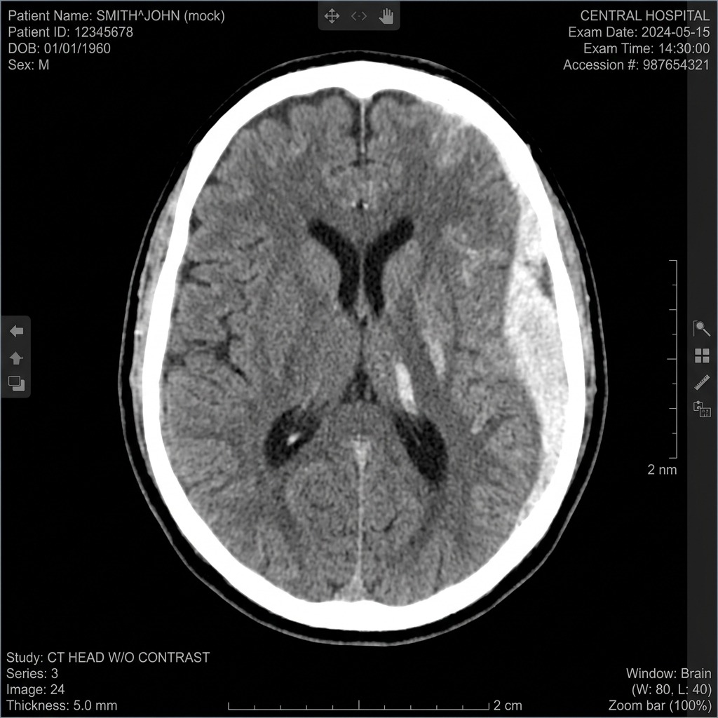
Acute Subdural Hematoma CT Head
Non-contrast head CT demonstrating an Acute Subdural Hematoma: Crescentic, concave hyperdensity tracking along the cerebral hemisphere that crosses cranial suture lines but is restricted by dural venous reflections (falx cerebri). Caused by tearing of bridging cortical veins. In contrast, epidural hematomas are biconvex (lens-shaped) and restricted by suture lines.
Intracranial Hemorrhage Differentiation
Clinical Matrix
Hemorrhage TypeVascular Source & MechanismNon-Contrast Head CT AppearanceClinical Features & Pitfalls
Epidural Hematoma (EDH)Middle meningeal artery rupture (pterion fracture)Hyperdense biconvex (lens-shaped) collection; does NOT cross suture linesLucid interval followed by rapid uncal herniation (ipsilateral blown pupil, contralateral hemiparesis)
Subdural Hematoma (SDH)Bridging cortical veins rupture (acceleration-deceleration)Crescent-shaped collection; crosses suture lines, bounded by dural reflectionsElderly patients, chronic alcoholics; insidious onset of headache and cognitive decline
Subarachnoid Hemorrhage (SAH)Ruptured saccular (berry) aneurysm or AVMHyperdense blood in basal cisterns, sylvian fissures, and sulciThunderclap headache, meningismus; LP shows xanthochromia; treat with Nimodipine
Intracerebral Hemorrhage (ICH)Charcot-Bouchard microaneurysms (chronic HTN)Hyperdense blood within brain parenchyma (basal ganglia, thalamus, pons)Focal deficit corresponding to neuroanatomy; target systolic BP 140 mmHg with IV nicardipine/labetalol
Board Trap — Status Epilepticus Treatment Escalation Timeline
High-Yield Board Trap & Alert
Status epilepticus is defined as ≥ 5 minutes of continuous seizure activity or ≥ 2 discrete seizures without complete return to consciousness between them. First-line therapy is immediate IV Lorazepam (4 mg IV bolus) or IM Midazolam (10 mg IM if no IV access). If seizures continue past 10–15 minutes, proceed immediately to second-line IV non-sedating antiepileptics: Levetiracetam (60 mg/kg), Fosphenytoin (20 mg PE/kg), or Valproate sodium (40 mg/kg). If seizures persist beyond 30 minutes (refractory status epilepticus), endotracheal intubation and continuous anesthetic infusions (Propofol, Midazolam, or Ketamine) with continuous EEG monitoring are mandatory.

Chapter 5 Quick-Check Self-Assessment

Test your clinical reasoning before moving to the next chapter
3 Vignettes
Board Vignette #1 Single Best Answer
A 23-year-old male is struck in the right temporal region with a baseball. He sustains a brief loss of consciousness for 30 seconds, awakens fully oriented (lucid interval), but 2 hours later becomes stuporous with a dilated, non-reactive right pupil and left hemiparesis. What vascular structure has ruptured?
Board Vignette #2 Single Best Answer
A 44-year-old female presents to the emergency department with the sudden onset of the 'worst headache of my life' that peaked instantaneously within seconds while lifting groceries. She has mild neck stiffness and photophobia. Vital signs are normal. Non-contrast head CT performed 4 hours after headache onset is completely normal. Which of the following is the most appropriate next step in management?
Board Vignette #3 Single Best Answer
A 32-year-old male with epilepsy is brought in by EMS with ongoing continuous generalized tonic-clonic convulsions lasting 12 minutes. Two doses of IV lorazepam (4 mg each) have failed to stop the seizure. What is the next most appropriate pharmacotherapy?
Estimated study time: 14 min
Chapter 6 • Gastrointestinal & Abdominal Emergencies
13 min TOC

Acute Abdominal & Gastrointestinal Emergencies

Upper vs. Lower GI Bleeding, Acute Appendicitis, Bowel Obstruction, Mesenteric Ischemia, and Pancreatitis

Clinical Overview & Board Focus

The acute abdomen encompasses inflammatory, obstructive, vascular, and hemorrhagic pathology requiring rapid decision-making regarding conservative management vs. emergent surgical exploration. Board exams test physical exam signs, targeted radiological modalities, and hemodynamic resuscitation algorithms.

6.1 The Acute Abdomen & Emergent Vascular Catastrophes

Acute appendicitis is the most common emergency surgical condition. Obstruction of the appendiceal lumen by a fecalith or lymphoid hyperplasia leads to luminal distention, visceral pain in the periumbilical region (T10 dermatome), followed by transmural inflammation and localized parietal peritoneal irritation in the right lower quadrant (McBurney's point). Hallmark exam signs include Rovsing's sign (RLQ pain on LLQ palpation), Psoas sign (pain on passive right hip extension), and Obturator sign (pain on passive internal rotation of the flexed right hip). In non-pregnant adults, contrast-enhanced CT of the abdomen/pelvis is the diagnostic gold standard; in children and pregnant patients, graded compression ultrasonography or pelvic MRI is preferred to avoid ionizing radiation.

Acute Mesenteric Ischemia is a critical vascular emergency classically characterized by 'pain out of proportion to physical examination' in an elderly patient with underlying atrial fibrillation, peripheral vascular disease, or recent myocardial infarction. The most common etiology (50%) is acute embolism from the left atrium to the superior mesenteric artery (SMA). Initial abdominal exam is deceptively benign without peritoneal signs; however, progression to transmural bowel infarction produces peritoneal signs, hematochezia, severe lactic acidosis, and sepsis. CT mesenteric angiography is the diagnostic test of choice (revealing arterial occlusion, bowel wall thickening, or pneumatosis intestinalis). Emergent surgical embolectomy, revascularization, and resection of necrotic bowel are life-saving.

Acute Upper Gastrointestinal Bleeding (UGIB) originates proximal to the ligament of Treitz, presenting as hematemesis ('coffee-ground' or bright red) or melena. Resuscitation requires two large-bore IVs (16-gauge or 18-gauge), rapid crystalloid boluses, and restrictive blood transfusion targeting hemoglobin ≥ 7 g/dL (or ≥ 8 g/dL in coronary artery disease). In suspected variceal bleeding (cirrhosis, stigmata of chronic liver disease), IV Octreotide (50 mcg bolus followed by 50 mcg/hr infusion) reduces portal venous pressure, and prophylactic Ceftriaxone (1 g IV daily) significantly decreases bacterial peritonitis, rebleeding, and mortality. Emergent upper endoscopy (EGD) within 24 hours provides definitive diagnosis and endoscopic band ligation.

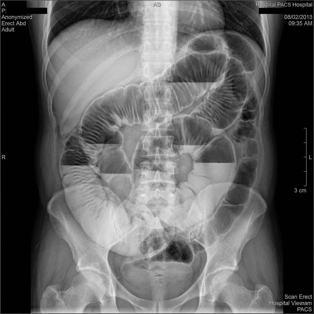
Small Bowel Obstruction Abdominal Radiograph
Upright abdominal radiograph demonstrating multiple dilated loops of small bowel with prominent air-fluid levels in a 'stepladder' distribution and absence of colonic gas, diagnostic of Mechanical Small Bowel Obstruction (SBO). Prior surgical adhesions represent the #1 etiology (>60%).
Acute Abdominal Emergencies — Clinical Summary
Clinical Matrix
EntityKey Risk Factors / EtiologyDiagnostic Modality of ChoiceFirst-Line Clinical Management
Acute CholecystitisGallstone impaction in cystic ductRUQ Ultrasound (sonographic Murphy sign, wall thickening > 3 mm, pericholecystic fluid)IV fluids, NPO, IV Ceftriaxone + Metronidazole, early Laparoscopic Cholecystectomy (< 72 hr)
Acute CholangitisCholedocholithiasis causing infected biliary stasisRUQ Ultrasound / CT abdomen; Reynolds' pentad (Charcot triad + shock + AMS)Emergent biliary decompression via ERCP + broad-spectrum IV antibiotics
Acute PancreatitisGallstones (40%), Alcohol (35%), Hypertriglyceridemia (> 1000 mg/dL)Lipase ≥ 3× upper limit of normal; CT not needed if lipase + classic epigastric pain presentAggressive IV Lactated Ringer's resuscitation, multimodal analgesia, early enteral feeding
Small Bowel Obstruction (SBO)Prior abdominal surgery (adhesions #1), incarcerated herniaAbdominal X-ray (dilated loops > 3 cm, air-fluid levels) or CT abdomen/pelvis with IV contrastNG tube decompression, bowel rest, IV fluids; emergent laparotomy if strangulated/perforated
COMLEX Clinical Integration — Viscerosomatic Collateral Reflexes in GI Pathology
COMLEX & OPP Board Integration
Abdominal viscera project characteristic viscerosomatic reflexes to the thoracic and lumbar paraspinal musculature via sympathetic splanchnic pathways: Stomach/Liver/Gallbladder project to T5–T9 (greater splanchnic nerve, celiac ganglion); Small Intestine and Ascending Colon project to T9–T11 (lesser splanchnic nerve, superior mesenteric ganglion); Descending Colon and Pelvic Organs project to T12–L2 (least and lumbar splanchnics, inferior mesenteric ganglion). Chapman's reflex point for the gallbladder is located in the 6th right intercostal space midclavicular line; the appendix Chapman's point is at the tip of the 12th right rib. Collateral ganglion release (celiac, SMG, IMG) and gentle paraspinal inhibition relieve splanchnic vasospasm and facilitate postoperative ileus resolution.

Chapter 6 Quick-Check Self-Assessment

Test your clinical reasoning before moving to the next chapter

3 Vignettes
Board Vignette #1 Single Best Answer
A 74-year-old male with chronic atrial fibrillation presents with sudden, excruciating diffuse abdominal pain. On physical exam, his abdomen is soft and non-distended with mild vague tenderness but no guarding, rigidity, or rebound tenderness. Serum lactate is 4.8 mmol/L. What is the most appropriate next diagnostic step?
Board Vignette #2 Single Best Answer
A 62-year-old male with a long history of chronic osteoarthritis treated with high-dose daily naproxen presents with acute, excruciating epigastric pain that began abruptly 2 hours ago. He is lying completely motionless on the stretcher and groans when the bed is bumped. Abdominal examination reveals board-like rigidity, absent bowel sounds, and diffuse severe rebound tenderness. Upright chest radiography reveals a thin crescent of air beneath the right hemidiaphragm. Which of the following is the most appropriate next step in management?
Board Vignette #3 Single Best Answer
A 72-year-old female presents with 3 days of progressive obstipation, abdominal distension, and colicky pain. Upright abdominal X-ray reveals an inverted 'U-shaped' dilated loop of colon with no haustra, pointing toward the right upper quadrant ('coffee bean sign'). What is the initial management of choice?
Estimated study time: 13 min
Chapter 7 • Environmental & Hypothermia
12 min TOC

Environmental Emergencies, Burns & Dysbarism

Accidental Hypothermia, Heat Exhaustion vs. Heat Stroke, Burn Resuscitation, and Electrical Injuries

Clinical Overview & Board Focus

Environmental illnesses occur when physiological homeostatic thermoregulation is overwhelmed by ambient extremes, immersion, burns, or submersion. Board examinations prioritize the distinction between heat exhaustion and heat stroke, rewarming protocols and arrhythmia management in severe hypothermia, and fluid resuscitation formulas for severe thermal burns.

7.1 Hypothermia, Heat Emergencies & Major Thermal Resuscitation

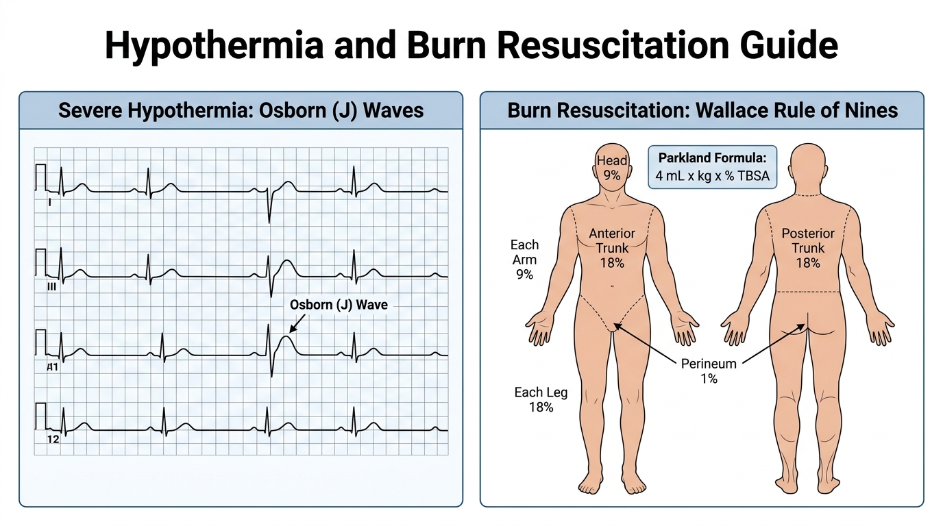
Accidental hypothermia is defined as an involuntary drop in core body temperature < 35°C (95°F). It is stratified into Mild (32–35°C: vigorous shivering, ataxia, dysarthria), Moderate (28–32°C: cessation of shivering, hypoventilation, altered mental status, J-waves / Osborn waves on ECG), and Severe (< 28°C: comatose, extreme bradycardia or ventricular fibrillation, apparent rigor mortis). In severe hypothermia, the myocardium is exceedingly irritable; rough handling or unnecessary movement can trigger refractory ventricular fibrillation. If VF occurs, defibrillation and ACLS medications are typically ineffective until the patient is rewarmed > 30°C. Active internal rewarming methods include warmed humidified O2, warmed IV crystalloids (40–42°C), thoracic or peritoneal lavage with warmed fluids, and extracorporeal membrane oxygenation (ECMO). A key board dictum: 'a patient is not dead until warm and dead' (core temp > 32°C with absent vitals).

Heat emergencies exist on a spectrum from Heat Exhaustion (core body temp < 40°C, intact mental status, profuse sweating, headache, nausea) to Heat Stroke (core body temp > 40°C [104°F] accompanied by profound central nervous system dysfunction: encephalopathy, delirium, seizures, or coma). Heat stroke is a medical emergency with high mortality from multiorgan failure and rhabdomyolysis. Immediate, aggressive evaporative or ice water immersion cooling is the treatment of choice, aiming to rapidly lower core temperature to < 39°C. Antipyretics (acetaminophen, aspirin) have NO role because hypothalamic temperature set-point is normal.

Thermal burns are quantified using the Rule of Nines: Head (9%), Entire Anterior Trunk (18%), Entire Posterior Trunk (18%), Each Upper Extremity (9%), Each Lower Extremity (18%), Perineum (1%). Fluid resuscitation for 2nd and 3rd degree burns > 20% Total Body Surface Area (TBSA) is calculated using the Parkland Formula: 4 mL × body weight (kg) × % TBSA burned, administering half of the total volume over the first 8 hours from the time of burn, and the remaining half over the subsequent 16 hours. Resuscitation is titrated to maintain a target urine output of 0.5–1.0 mL/kg/hr in adults.

Hypothermia Osborn J Waves and Burn Rule of Nines Resuscitation
Diagnostic and resuscitation guide for environmental emergencies: Left panel demonstrates pathognomonic Osborn (J) Waves (prominent positive deflection at the J-point / QRS-ST junction) in severe accidental hypothermia with sinus bradycardia. Right panel outlines the Wallace Rule of Nines Total Body Surface Area (TBSA) breakdown paired with the Parkland Formula (4 mL × kg × % TBSA) for major thermal burn crystalloid resuscitation.
Board Trap — Circumferential Full-Thickness Burns & Escharotomy
High-Yield Board Trap & Alert
Full-thickness (3rd-degree) burns form a rigid, leathery, non-elastic eschar. In circumferential extremity burns, ongoing tissue edema elevates sub-eschar pressure, producing a severe burn-induced compartment syndrome with loss of distal pulses. In circumferential thoracic burns, rigid eschar restricts chest wall expansion, leading to severe hypoventilation, high peak airway pressures, and hypoxia. Emergent bedside escharotomy (longitudinal incisions through the depth of the eschar into subcutaneous fat until edges separate) is life-saving and does not require general anesthesia because full-thickness burns destroy dermal nerve endings.

Chapter 7 Quick-Check Self-Assessment

Test your clinical reasoning before moving to the next chapter

3 Vignettes
Board Vignette #1 Single Best Answer
A 19-year-old military recruit collapses during basic training on a hot summer afternoon (ambient temperature 38°C). On arrival, he is delirious and obtunded. Rectal core temperature is 41.2°C (106.2°F). Vitals: BP 86/50 mmHg, HR 148 bpm. Skin is hot and anhidrotic. What is the single most urgent therapeutic intervention?
Board Vignette #2 Single Best Answer
An 82-year-old woman is brought to the ED during a summer heat wave after being found confused and agitated in her unairconditioned apartment. Her temperature is 40.8°C (105.4°F), blood pressure is 96/58 mmHg, heart rate is 124 bpm, and respirations are 26/min. Her skin is hot, flushed, and dry. Neurological exam reveals profound confusion and disorientation. Serum labs show AST 480 U/L, ALT 510 U/L, and BUN/Cr 38/2.1. Which of the following is the most appropriate immediate intervention?
Board Vignette #3 Single Best Answer
A 22-year-old military recruit collapses during a 10-mile march in 95°F heat. Core rectal temperature is 105.8°F (41.0°C). He is combative, delirious, and profusely diaphoretic with BP 86/52 mmHg. What is the most critical immediate intervention to reduce mortality?
Estimated study time: 12 min